")
Tie-2 Antibody (3A5): sc-293414
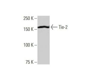
- Tie-2 Antibody (3A5) is a mouse monoclonal IgG2a κ Tie-2 antibody, cited in 11 publications, provided at 100 µg/ml
- raised against amino acids 66-185 representing partial recombinant protein of Tie-2 of human origin
- recommended for detection of Tie-2 of mouse, rat and human origin by WB, IP and ELISA
- m-IgG Fc BP-HRP, m-IgG2a BP-HRP and m-IgGκ BP-HRP are the preferred secondary detection reagents for Tie-2 Antibody (3A5) for WB applications. These reagents are now offered in bundles with Tie-2 Antibody (3A5) (see ordering information below).

Tie-2 Antibody (3A5) is a mouse monoclonal IgG2a kappa light chain antibody that detects Tie-2 protein of mouse, rat, and human origin by western blotting (WB), immunoprecipitation (IP), and enzyme-linked immunosorbent assay (ELISA). Anti-Tie-2 antibody (3A5) is available as the non-conjugated form, which is crucial for studying the role of receptor tyrosine kinases in various biological processes, particularly in the vascular system. Tie-2 (3A5) antibody recognizes Tie-2, also known as Tek, which is primarily located on the surface of endothelial cells and plays a vital role in angiogenesis and vascular stability. Tie-2 (3A5) monoclonal antibody enables research into the regulation of blood vessel formation and maintenance, as Tie-2 mediates signaling pathways that influence endothelial cell survival, proliferation, and migration. Dysregulation of Tie-2 signaling has been implicated in various pathological conditions, including cancer and cardiovascular diseases, making Tie-2 a significant target for therapeutic intervention. Anti-Tie-2 antibody (3A5) helps researchers study Tie-2′s interactions with several ligands, including angiopoietins, which are critical for promoting vascular integrity and remodeling. Tie-2 (3A5) monoclonal antibody provides valuable insights into the mechanisms of vascular development and potential implications of Tie-2 in disease states.
Alexa Fluor® is a trademark of Molecular Probes Inc., OR., USA
LI-COR® and Odyssey® are registered trademarks of LI-COR Biosciences
Tie-2 Antibody (3A5) References:
- The fms-like tyrosine kinase, a receptor for vascular endothelial growth factor. | de Vries, C., et al. 1992. Science. 255: 989-91. PMID: 1312256
- A novel endothelial cell surface receptor tyrosine kinase with extracellular epidermal growth factor homology domains. | Partanen, J., et al. 1992. Mol Cell Biol. 12: 1698-707. PMID: 1312667
- tek, a novel tyrosine kinase gene located on mouse chromosome 4, is expressed in endothelial cells and their presumptive precursors. | Dumont, DJ., et al. 1992. Oncogene. 7: 1471-80. PMID: 1630810
- Localization of Ang-1, -2, Tie-2, and VEGF expression at endothelial-pericyte interdigitation in rat angiogenesis. | Wakui, S., et al. 2006. Lab Invest. 86: 1172-84. PMID: 16969369
- Receptor tyrosine kinases: genetic evidence for their role in Drosophila and mouse development. | Pawson, T. and Bernstein, A. 1990. Trends Genet. 6: 350-6. PMID: 1965067
- Placental mRNA expression of angiopoietins (Ang)-1, Ang-2 and their receptor Tie-2 is altered in pregnancies complicated by preeclampsia. | Kappou, D., et al. 2014. Placenta. 35: 718-23. PMID: 25047691
- T11TS inhibits Angiopoietin-1/Tie-2 signaling, EGFR activation and Raf/MEK/ERK pathway in brain endothelial cells restraining angiogenesis in glioma model. | Bhattacharya, D., et al. 2015. Exp Mol Pathol. 98: 455-66. PMID: 25797371
- Lack of FADD in Tie-2 expressing cells causes RIPK3-mediated embryonic lethality. | Fan, C., et al. 2016. Cell Death Dis. 7: e2351. PMID: 27584790
- Expression and significance of B7-H3 and Tie-2 in the tumor vasculature of clear cell renal carcinoma. | Zhang, X., et al. 2017. Onco Targets Ther. 10: 5417-5424. PMID: 29180874
- Hyperoxia-induced S1P1 signaling reduced angiogenesis by suppression of TIE-2 leading to experimental bronchopulmonary dysplasia. | Sudhadevi, T., et al. 2021. Cell Biochem Biophys. 79: 561-573. PMID: 34176100
- The endothelial-specific receptor tyrosine kinase, tek, is a member of a new subfamily of receptors. | Dumont, DJ., et al. 1993. Oncogene. 8: 1293-301. PMID: 8386827
- Tie-1 and tie-2 define another class of putative receptor tyrosine kinase genes expressed in early embryonic vascular system. | Sato, TN., et al. 1993. Proc Natl Acad Sci U S A. 90: 9355-8. PMID: 8415706
Ordering Information
| Product Name | Catalog # | UNIT | Price | Qty | FAVORITES | |
Tie-2 Antibody (3A5) | sc-293414 | 100 µg/ml | $322.00 | |||
Tie-2 Antibody (3A5): m-IgG Fc BP-HRP Bundle | sc-537744 | 100 µg Ab; 10 µg BP | $361.00 | |||
Tie-2 Antibody (3A5): m-IgGκ BP-HRP Bundle | sc-535110 | 100 µg Ab; 40 µg BP | $361.00 | |||
Tie-2 Antibody (3A5): m-IgG2a BP-HRP Bundle | sc-548106 | 100 µg Ab; 10 µg BP | $361.00 |