")
: sc-393127. Western blot analysis of PAR-3 expression in ARPE-19 (A), SK-N-SH (B), RAW 264.7 (C), WEHI-231 (D), RPE-J (E) and KNRK (F) whole cell lysates.")
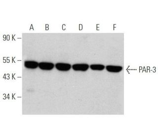
: sc-393127. Western blot analysis of PAR-3 expression in HL-60 (A), MEG-01 (B), K-562 (C), CCD-1064Sk (D), LADMAC (E) and M1 (F) whole cell lysates.")
: sc-393127. Western blot analysis of PAR-3 expression in NIH/3T3 whole cell lysate.")
PAR-3 Antibody (G-4): sc-393127
- PAR-3 Antibody (G-4) is a mouse monoclonal IgG1 κ PAR-3 antibody provided at 200 µg/ml
- raised against amino acids 1-103 of PAR-3 of human origin
- PAR-3 Antibody (G-4) is recommended for detection of PAR-3 of mouse, rat and human origin by WB, IP, IF and ELISA
- Anti-PAR-3 Antibody (G-4) is available conjugated to agarose for IP; HRP for WB, IHC(P) and ELISA; and to either phycoerythrin or FITC for IF, IHC(P) and FCM
- also available conjugated to Alexa Fluor® 488, Alexa Fluor® 546, Alexa Fluor® 594 or Alexa Fluor® 647 for WB (RGB), IF, IHC(P) and FCM, and for use with RGB fluorescent imaging systems, such as iBright™ FL1000, FluorChem™, Typhoon, Azure and other comparable systems
- also available conjugated to Alexa Fluor® 680 or Alexa Fluor® 790 for WB (NIR), IF and FCM; for use with Near-Infrared (NIR) detection systems, such as LI-COR®Odyssey®, iBright™ FL1000, FluorChem™, Typhoon, Azure and other comparable systems
- m-IgG Fc BP-HRP, m-IgG1 BP-HRP and m-IgGκ BP-HRP are the preferred secondary detection reagents for PAR-3 Antibody (G-4) for WB applications. These reagents are now offered in bundles with PAR-3 Antibody (G-4) (see ordering information below).

QUICK LINKS
PAR-3 Antibody (G-4) is a mouse monoclonal IgG1 kappa light chain antibody that detects PAR-3 protein of mouse, rat, and human origin by western blotting (WB), immunoprecipitation (IP), immunofluorescence (IF), and enzyme-linked immunosorbent assay (ELISA). PAR-3 Antibody (G-4) is available in both non-conjugated and various conjugated forms, including agarose, horseradish peroxidase (HRP), phycoerythrin (PE), fluorescein isothiocyanate (FITC), and multiple Alexa Fluor® conjugates. PAR-3, also known as protease-activated receptor 3, plays a crucial role in signaling pathways activated by thrombin, a serine protease essential for blood coagulation and platelet aggregation. Upon activation, PAR-3 influences cellular responses such as mitogenesis and thrombosis, highlighting PAR-3′s importance in vascular biology and hemostasis. PAR-3 is primarily located in the cell membrane, where PAR-3 functions as a G protein-coupled receptor, facilitating communication between extracellular signals and intracellular responses. This localization is vital for PAR-3′s role in mediating effects of thrombin and other proteases, which can lead to significant physiological outcomes, including inflammation and tissue repair. Understanding PAR-3′s function and location is essential for elucidating PAR-3′s contributions to various pathophysiological conditions, including cardiovascular diseases and inflammatory responses.
Alexa Fluor® is a trademark of Molecular Probes Inc., OR., USA
LI-COR® and Odyssey® are registered trademarks of LI-COR Biosciences
PAR-3 Antibody (G-4) References:
- The Par-3 NTD adopts a PB1-like structure required for Par-3 oligomerization and membrane localization. | Feng, W., et al. 2007. EMBO J. 26: 2786-96. PMID: 17476308
- Phosphoinositide binding by par-3 involved in par-3 localization. | Horikoshi, Y., et al. 2011. Cell Struct Funct. 36: 97-102. PMID: 21467691
- Loss of Par3 promotes lung adenocarcinoma metastasis through 14-3-3ζ protein. | Song, T., et al. 2016. Oncotarget. 7: 64260-64273. PMID: 27588399
- Flow cytometry analysis reveals different activation profiles of thrombin- or TRAP-stimulated platelets in db/db mice. The regulatory role of PAR-3. | Kassassir, H., et al. 2017. Blood Cells Mol Dis. 65: 16-22. PMID: 28460264
- PAR-3 controls endothelial planar polarity and vascular inflammation under laminar flow. | Hikita, T., et al. 2018. EMBO Rep. 19: PMID: 30018153
- A Conserved PDZ-Binding Motif in aPKC Interacts with Par-3 and Mediates Cortical Polarity. | Holly, RW., et al. 2020. Curr Biol. 30: 893-898.e5. PMID: 32084408
- Polarized endosome dynamics engage cytoplasmic Par-3 that recruits dynein during asymmetric cell division. | Zhao, X., et al. 2021. Sci Adv. 7: PMID: 34117063
- A particle size threshold governs diffusion and segregation of PAR-3 during cell polarization. | Chang, Y. and Dickinson, DJ. 2022. Cell Rep. 39: 110652. PMID: 35417695
- The Roles of Par3, Par6, and aPKC Polarity Proteins in Normal Neurodevelopment and in Neurodegenerative and Neuropsychiatric Disorders. | Zhang, L. and Wei, X. 2022. J Neurosci. 42: 4774-4793. PMID: 35705493
- EPH receptor tyrosine kinases phosphorylate the PAR-3 scaffold protein to modulate downstream signaling networks. | Banerjee, SL., et al. 2022. Cell Rep. 40: 111031. PMID: 35793621
Ordering Information
| Product Name | Catalog # | UNIT | Price | Qty | FAVORITES | |
PAR-3 Antibody (G-4) | sc-393127 | 200 µg/ml | $322.00 | |||
PAR-3 Antibody (G-4): m-IgG Fc BP-HRP Bundle | sc-530363 | 200 µg Ab; 10 µg BP | $361.00 | |||
PAR-3 Antibody (G-4): m-IgGκ BP-HRP Bundle | sc-523816 | 200 µg Ab, 40 µg BP | $361.00 | |||
PAR-3 Antibody (G-4): m-IgG1 BP-HRP Bundle | sc-544014 | 200 µg Ab; 20 µg BP | $361.00 | |||
PAR-3 Antibody (G-4) AC | sc-393127 AC | 500 µg/ml, 25% agarose | $424.00 | |||
PAR-3 Antibody (G-4) HRP | sc-393127 HRP | 200 µg/ml | $322.00 | |||
PAR-3 Antibody (G-4) FITC | sc-393127 FITC | 200 µg/ml | $336.00 | |||
PAR-3 Antibody (G-4) PE | sc-393127 PE | 200 µg/ml | $349.00 | |||
PAR-3 Antibody (G-4) Alexa Fluor® 488 | sc-393127 AF488 | 200 µg/ml | $364.00 | |||
PAR-3 Antibody (G-4) Alexa Fluor® 546 | sc-393127 AF546 | 200 µg/ml | $364.00 | |||
PAR-3 Antibody (G-4) Alexa Fluor® 594 | sc-393127 AF594 | 200 µg/ml | $364.00 | |||
PAR-3 Antibody (G-4) Alexa Fluor® 647 | sc-393127 AF647 | 200 µg/ml | $364.00 | |||
PAR-3 Antibody (G-4) Alexa Fluor® 680 | sc-393127 AF680 | 200 µg/ml | $364.00 | |||
PAR-3 Antibody (G-4) Alexa Fluor® 790 | sc-393127 AF790 | 200 µg/ml | $364.00 |