
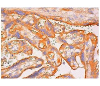
IDO Antibody (E-1): sc-376413
- IDO Antibody (E-1) is a mouse monoclonal IgG2b κ, cited in 2 publications, provided at 200 µg/ml
- specific for an epitope mapping between amino acids 361-399 near the C-terminus of IDO of mouse origin
- Anti-IDO Antibody (E-1) is recommended for detection of IDO of mouse, rat and human origin by WB, IP, IF, IHC(P) and ELISA
- Anti-IDO Antibody (E-1) is available conjugated to agarose for IP; HRP for WB, IHC(P) and ELISA; and to either phycoerythrin or FITC for IF, IHC(P) and FCM
- also available conjugated to Alexa Fluor® 488, Alexa Fluor® 546, Alexa Fluor® 594 or Alexa Fluor® 647 for WB (RGB), IF, IHC(P) and FCM, and for use with RGB fluorescent imaging systems, such as iBright™ FL1000, FluorChem™, Typhoon, Azure and other comparable systems
- also available conjugated to Alexa Fluor® 680 or Alexa Fluor® 790 for WB (NIR), IF and FCM; for use with Near-Infrared (NIR) detection systems, such as LI-COR®Odyssey®, iBright™ FL1000, FluorChem™, Typhoon, Azure and other comparable systems
- m-IgG Fc BP-HRP and m-IgG2b BP-HRP are the preferred secondary detection reagents for IDO Antibody (E-1) for WB and IHC(P) applications. These reagents are now offered in bundles with IDO Antibody (E-1) (see ordering information below).

QUICK LINKS
IDO Antibody (E-1) is a mouse monoclonal IgG2b kappa light chain antibody that detects IDO protein from mouse, rat, and human origin by western blotting (WB), immunoprecipitation (IP), immunofluorescence (IF), immunohistochemistry with paraffin-embedded sections (IHCP), and enzyme-linked immunosorbent assay (ELISA). Anti-IDO antibody (E-1) is available in both non-conjugated and various conjugated forms, including agarose, horseradish peroxidase (HRP), phycoerythrin (PE), fluorescein isothiocyanate (FITC), and multiple Alexa Fluor® conjugates. Indoleamine 2,3-dioxygenase (IDO) plays a crucial role in immune regulation by catalyzing the degradation of the essential amino acid L-tryptophan into N-formylkynurenine, a process particularly important in immune tolerance and response. IDO is induced by interferon-gamma and modulates immunological responses, protecting allogeneic concepti from maternal immune rejection. IDO activity can inhibit T cell proliferation, as demonstrated in studies where IDO-transfected cells co-cultured with T cells resulted in reduced T cell activity. IDO presence in dendritic cells suppresses T cell responses, highlighting IDO′s significance in maintaining immune homeostasis. Human IDO gene is located on chromosome 8p12-p11, and understanding IDO function and interactions remains vital for developing therapeutic strategies aimed at manipulating immune responses, particularly in cancer and transplantation settings.
Alexa Fluor® is a trademark of Molecular Probes Inc., OR., USA
LI-COR® and Odyssey® are registered trademarks of LI-COR Biosciences
Ordering Information
| Product Name | Catalog # | UNIT | Price | Qty | FAVORITES | |
IDO Antibody (E-1) | sc-376413 | 200 µg/ml | $322.00 | |||
IDO Antibody (E-1): m-IgG Fc BP-HRP Bundle | sc-526027 | 200 µg Ab; 10 µg BP | $361.00 | |||
IDO Antibody (E-1): m-IgG2b BP-HRP Bundle | sc-549663 | 200 µg Ab; 10 µg BP | $361.00 | |||
IDO Antibody (E-1) AC | sc-376413 AC | 500 µg/ml, 25% agarose | $424.00 | |||
IDO Antibody (E-1) HRP | sc-376413 HRP | 200 µg/ml | $322.00 | |||
IDO Antibody (E-1) FITC | sc-376413 FITC | 200 µg/ml | $336.00 | |||
IDO Antibody (E-1) PE | sc-376413 PE | 200 µg/ml | $349.00 | |||
IDO Antibody (E-1) Alexa Fluor® 488 | sc-376413 AF488 | 200 µg/ml | $364.00 | |||
IDO Antibody (E-1) Alexa Fluor® 546 | sc-376413 AF546 | 200 µg/ml | $364.00 | |||
IDO Antibody (E-1) Alexa Fluor® 594 | sc-376413 AF594 | 200 µg/ml | $364.00 | |||
IDO Antibody (E-1) Alexa Fluor® 647 | sc-376413 AF647 | 200 µg/ml | $364.00 | |||
IDO Antibody (E-1) Alexa Fluor® 680 | sc-376413 AF680 | 200 µg/ml | $364.00 | |||
IDO Antibody (E-1) Alexa Fluor® 790 | sc-376413 AF790 | 200 µg/ml | $364.00 | |||
IDO (E-1) Neutralizing Peptide | sc-376413 P | 100 µg/0.5 ml | $69.00 |