")
: sc-52658. Análisis por Western blot de la expresión de COL2A1 en lisado de células enteras CCD-1064Sk.")
: sc-52658. Análisis de Western blot de la expresión de COL2A1 en lisados de células enteras de HISM (A) y CCD-1064Sk (B).")
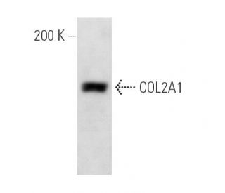
: sc-52658. Análisis por Western blot de la expresión de COL2A1 en lisado de células enteras de HISM.")
COL2A1 Anticuerpo (M2139): sc-52658
- COL2A1 Anticuerpo M2139 es un monoclonal de ratón IgG2b κ COL2A1 Anticuerpo, ver las 172 publicaciones, proporcionado como 100 µg/ml
- planteada frente al epítopo J1 del colágeno II (posición 551-564 de la triple hélice) de origen mouse.
- recomendado para detectar Collagen Type II de mouse, rat, human y Arabadopsis origen, mediante WB, IP, IF y IHC(P); también reactivo con otras especies, incluyendo bovine
- m-IgG Fc BP-HRP y 2b BP-HRP">m-IgG2b BP-HRP son los reactivos de detección secundarios preferidos para COL2A1 Anticuerpo (M2139) para aplicaciones WB e IHC(P). Estos reactivos se ofrecen ahora en paquetes con COL2A1 Anticuerpo (M2139)(véase la información de pedido más abajo).

ENLACES RÁPIDOS
El anticuerpo COL2A1 (M2139) es un anticuerpo monoclonal IgG2b κ de ratón COL2A1 (también designado como anticuerpo de la cadena alfa 1 del colágeno tipo II) que detecta la proteína colágeno tipo II de origen de ratón, rata, humano y Arabadopsis por WB, IP, IF e IHC(P). El anticuerpo COL2A1 (M2139) está disponible como anticuerpo anti-COL2A1 no conjugado. La extensa familia de productos génicos COL (colágenos) está compuesta por varios tipos de cadenas, incluyendo colágenos intersticiales formadores de fibrillas (tipos I, II, III y V) y colágenos de membrana basal (tipo IV), cada tipo conteniendo múltiples isoformas. Los colágenos son proteínas fibrosas de matriz extracelular con gran resistencia a la tracción y son los principales componentes del tejido conjuntivo, como los tendones y los cartílagos. Todos los colágenos contienen un dominio de triple hélice y con frecuencia muestran autoasociación lateral para formar tejidos conectivos complejos. Varios colágenos también desempeñan un papel en la adhesión celular, importante para mantener la arquitectura y la función normales de los tejidos. En el cartílago, el Colágeno Tipo II constituye la mayor parte de la fibrilla. La sensibilización con Colágeno Tipo II induce una poliartritis erosiva en ratas, ratones y primates superiores que puede parecerse a la artritis reumatoide y a la policrondritis recidivante.
Alexa Fluor® es una marca registrada de Molecular Probes Inc., OR., USA
REIVEW LI-COR® y Odyssey® son marcas registradas de LI-COR Biosciences.
COL2A1 Anticuerpo (M2139) Referencias:
- El rastreo del linaje de Col2a1 revela que el menisco de la articulación de la rodilla tiene un origen celular complejo. | Hyde, G., et al. 2008. J Anat. 213: 531-8. PMID: 19014360
- El deterioro esquelético en la displasia espondiloepifisaria relacionada con COL2A1 se produce antes de la osteoartritis. | Rolvien, T., et al. 2020. Osteoarthritis Cartilage. 28: 334-343. PMID: 31958497
- Nuevas variantes en COL2A1 causantes de displasia espondiloepifisaria congénita poco frecuente. | Zheng, WB., et al. 2020. Mol Genet Genomic Med. 8: e1139. PMID: 31972903
- Una nueva mutación intrónica profunda de COL2A1 en una familia con síndrome de Stickler de alta miopía/sólo ocular de aparición temprana. | Sun, W., et al. 2020. Ophthalmic Physiol Opt. 40: 281-288. PMID: 32196734
- COL2A1 es un nuevo biomarcador de células repobladoras de tumores de melanoma. | Talluri, B., et al. 2020. Biomedicines. 8: PMID: 32962144
- La secuenciación del exoma reveló mutaciones en USP9X y COL2A1 en una familia numerosa con displasia epifisaria múltiple. | Luo, ZJ., et al. 2022. Bone. 163: 116508. PMID: 35907616
- La complejidad molecular de las variantes de empalme de COL2A1 y su importancia en la gravedad del fenotipo. | Viakhireva, I., et al. 2024. Bone. 181: 117013. PMID: 38246255
- Adhesión celular a matrices colágenas. | McCarthy, JB., et al. 1996. Biopolymers. 40: 371-81. PMID: 8765608
- Tolerancia nasal a epítopos dominantes y subdominantes del colágeno tipo II y protección contra la artritis inducida por colágeno. | Staines, NA., et al. 1997. Biochem Soc Trans. 25: 661-4. PMID: 9191176
- Colágenos versátiles en invertebrados. | Engel, J. 1997. Science. 277: 1785-6. PMID: 9324767
- Artritis inducida por colágeno, un modelo animal de autoinmunidad. | Myers, LK., et al. 1997. Life Sci. 61: 1861-78. PMID: 9364191
- Los colágenos del cartílago: revisión de su estructura, organización y papel en la patogénesis de la artritis experimental en animales y en la enfermedad reumática humana. | Cremer, MA., et al. 1998. J Mol Med (Berl). 76: 275-88. PMID: 9535561
Información sobre pedidos
| Nombre del producto | Número de catálogo | UNIDAD | Precio | CANTIDAD | Favoritos | |
COL2A1 Anticuerpo (M2139) | sc-52658 | 100 µg/ml | $322.00 | |||
Paquete de COL2A1 (M2139): m-IgG Fc BP-HRP | sc-538958 | 100 µg Ab; 10 µg BP | $361.00 | |||
Paquete de COL2A1 (M2139): m-IgG2b BP-HRP | sc-549724 | 100 µg Ab; 10 µg BP | $361.00 |