")
YTHDF3 Antibody (F-2): sc-377119
- YTHDF3 Antibody (F-2) is a mouse monoclonal IgG2a κ YTHDF3 antibody, cited in 23 publications, provided at 200 µg/ml
- specific for an epitope mapping between amino acids 85-119 within an internal region of YTHDF3 of human origin
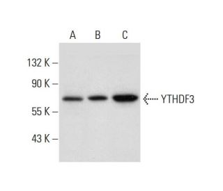
- YTHDF3 Antibody (F-2) is recommended for detection of YTHDF3 of mouse, rat, human and origin by WB, IP, IF, IHC(P) and ELISA; also reactive with additional species, including and equine, canine, bovine, porcine and avian
- Anti-YTHDF3 Antibody (F-2) is available conjugated to agarose for IP; HRP for WB, IHC(P) and ELISA; and to either phycoerythrin or FITC for IF, IHC(P) and FCM
- also available conjugated to Alexa Fluor® 488, Alexa Fluor® 546, Alexa Fluor® 594 or Alexa Fluor® 647 for WB (RGB), IF, IHC(P) and FCM, and for use with RGB fluorescent imaging systems, such as iBright™ FL1000, FluorChem™, Typhoon, Azure and other comparable systems
- also available conjugated to Alexa Fluor® 680 or Alexa Fluor® 790 for WB (NIR), IF and FCM; for use with Near-Infrared (NIR) detection systems, such as LI-COR®Odyssey®, iBright™ FL1000, FluorChem™, Typhoon, Azure and other comparable systems
- m-IgG Fc BP-HRP, m-IgG2a BP-HRP and m-IgGκ BP-HRP are the preferred secondary detection reagents for YTHDF3 Antibody (F-2) for WB and IHC(P) applications. These reagents are now offered in bundles with YTHDF3 Antibody (F-2) (see ordering information below).

QUICK LINKS
YTHDF3 Antibody (F-2) is a mouse monoclonal IgG2a kappa light chain antibody that detects YTHDF3 protein of mouse, rat, and human origin by western blotting (WB), immunoprecipitation (IP), immunofluorescence (IF), immunohistochemistry with paraffin-embedded sections (IHCP), and enzyme-linked immunosorbent assay (ELISA). anti-YTHDF3 antibody (F-2) is available in both non-conjugated and various conjugated forms, including agarose, horseradish peroxidase (HRP), phycoerythrin (PE), fluorescein isothiocyanate (FITC), and multiple Alexa Fluor® conjugates. YTHDF3, consisting of 585 amino acids, plays a crucial role in RNA metabolism, particularly in regulating mRNA stability and translation. YTHDF3 interacts with N6-methyladenosine (m6A) modified RNA, which is essential for various cellular processes, including gene expression and cellular response to stress. YTHDF3′s ability to bind to m6A-modified transcripts highlights YTHDF3′s importance in post-transcriptional regulation, influencing processes such as cell differentiation and proliferation. YTHDF3 is encoded on chromosome 8, a region associated with several genetic disorders, including certain leukemias and psychiatric conditions. Understanding YTHDF3′s function and interactions with m6A-modified RNA can provide insights into YTHDF3′s role in health and disease, making YTHDF3 monoclonal antibody (F-2) a valuable tool for researchers studying RNA biology and related pathways.
Alexa Fluor® is a trademark of Molecular Probes Inc., OR., USA
LI-COR® and Odyssey® are registered trademarks of LI-COR Biosciences
YTHDF3 Antibody (F-2) References:
- Chromosomes 8 and 10 workshop. | Wildenauer, DB. and Schwab, SG. 1999. Am J Med Genet. 88: 239-43. PMID: 10374738
- Preferential expression of an intact WRN gene in Werner syndrome cell lines in which a normal chromosome 8 has been introduced. | Kashino, G., et al. 2001. Biochem Biophys Res Commun. 289: 111-5. PMID: 11708785
- YTH: a new domain in nuclear proteins. | Stoilov, P., et al. 2002. Trends Biochem Sci. 27: 495-7. PMID: 12368078
- Combined analysis from eleven linkage studies of bipolar disorder provides strong evidence of susceptibility loci on chromosomes 6q and 8q. | McQueen, MB., et al. 2005. Am J Hum Genet. 77: 582-95. PMID: 16175504
- DNA sequence and analysis of human chromosome 8. | Nusbaum, C., et al. 2006. Nature. 439: 331-5. PMID: 16421571
- Epigenetic inactivation of the premature aging Werner syndrome gene in human cancer. | Agrelo, R., et al. 2006. Proc Natl Acad Sci U S A. 103: 8822-7. PMID: 16723399
- Non-Hodgkin's lymphomas with Burkitt-like cells are associated with c-Myc amplification and poor prognosis. | Mossafa, H., et al. 2006. Leuk Lymphoma. 47: 1885-93. PMID: 17065002
- Determining the DUF55-domain structure of human thymocyte nuclear protein 1 from crystals partially twinned by tetartohedry. | Yu, F., et al. 2009. Acta Crystallogr D Biol Crystallogr. 65: 212-9. PMID: 19237743
Ordering Information
| Product Name | Catalog # | UNIT | Price | Qty | FAVORITES | |
YTHDF3 Antibody (F-2) | sc-377119 | 200 µg/ml | $322.00 | |||
YTHDF3 Antibody (F-2): m-IgG Fc BP-HRP Bundle | sc-529978 | 200 µg Ab; 10 µg BP | $361.00 | |||
YTHDF3 Antibody (F-2): m-IgGκ BP-HRP Bundle | sc-523196 | 200 µg Ab, 40 µg BP | $361.00 | |||
YTHDF3 Antibody (F-2): m-IgG2a BP-HRP Bundle | sc-547412 | 200 µg Ab; 10 µg BP | $361.00 | |||
YTHDF3 Antibody (F-2) AC | sc-377119 AC | 500 µg/ml, 25% agarose | $424.00 | |||
YTHDF3 Antibody (F-2) HRP | sc-377119 HRP | 200 µg/ml | $322.00 | |||
YTHDF3 Antibody (F-2) FITC | sc-377119 FITC | 200 µg/ml | $336.00 | |||
YTHDF3 Antibody (F-2) PE | sc-377119 PE | 200 µg/ml | $349.00 | |||
YTHDF3 Antibody (F-2) Alexa Fluor® 488 | sc-377119 AF488 | 200 µg/ml | $364.00 | |||
YTHDF3 Antibody (F-2) Alexa Fluor® 546 | sc-377119 AF546 | 200 µg/ml | $364.00 | |||
YTHDF3 Antibody (F-2) Alexa Fluor® 594 | sc-377119 AF594 | 200 µg/ml | $364.00 | |||
YTHDF3 Antibody (F-2) Alexa Fluor® 647 | sc-377119 AF647 | 200 µg/ml | $364.00 | |||
YTHDF3 Antibody (F-2) Alexa Fluor® 680 | sc-377119 AF680 | 200 µg/ml | $364.00 | |||
YTHDF3 Antibody (F-2) Alexa Fluor® 790 | sc-377119 AF790 | 200 µg/ml | $364.00 | |||
YTHDF3 (F-2) Neutralizing Peptide | sc-377119 P | 100 µg/0.5 ml | $69.00 |