
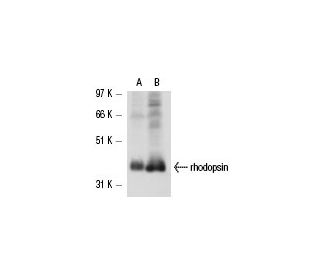


rhodopsin Antibody (C7): sc-53991
- rhodopsin Antibody (C7) is a mouse monoclonal IgG1 κ, cited in 2 publications, provided at 200 µg/ml
- raised against rhodopsin of bovine origin
- recommended for detection of of mouse, rat and bovine origin by WB, IP and IF
- See rhodopsin (1D4): sc-57432 for rhodopsin antibody conjugates, including AC, HRP, FITC, PE, Alexa Fluor® 488, 594, 647, 680 and 790.
- m-IgG Fc BP-HRP, m-IgG1 BP-HRP and m-IgGκ BP-HRP are the preferred secondary detection reagents for rhodopsin Antibody (C7) for WB applications. These reagents are now offered in bundles with rhodopsin Antibody (C7) (see ordering information below).

QUICK LINKS
SEE ALSO...
Rhodopsin Antibody (C7) is a mouse monoclonal IgG1 antibody that detects rhodopsin in mouse, rat, and bovine samples through applications such as western blotting (WB), immunoprecipitation (IP), and immunofluorescence (IF). Rhodopsin is a crucial photoreceptor protein located in the outer segments of rod photoreceptor cells in the retina, where rhodopsin plays a vital role in the visual transduction pathway. Anti-rhodopsin antibody (C7) is essential for studying protein conversion of light into electrical signals, enabling vision in low-light conditions. The structure of rhodopsin is characterized by seven transmembrane α-helices, which form a binding pocket for the chromophore 11-cis-retinal. Upon photon absorption, 11-cis-retinal undergoes isomerization to all-trans-retinal, triggering a conformational change in rhodopsin that activates the associated G protein, transducin. This activation initiates a cascade of biochemical events that ultimately lead to the perception of light. Mutations in the rhodopsin gene can disrupt this process, resulting in conditions such as retinitis pigmentosa, which can lead to progressive vision loss. Rhodopsin monoclonal antibody (C7) is an invaluable tool for researchers studying the mechanisms of vision and the pathophysiology of retinal diseases.
Alexa Fluor® is a trademark of Molecular Probes Inc., OR., USA
LI-COR® and Odyssey® are registered trademarks of LI-COR Biosciences
rhodopsin Antibody (C7) References:
- Structural basis for sensory rhodopsin function. | Pebay-Peyroula, E., et al. 2002. Biochim Biophys Acta. 1565: 196-205. PMID: 12409195
- Recoverin and rhodopsin kinase. | Chen, CK. 2002. Adv Exp Med Biol. 514: 101-7. PMID: 12596917
- Rhodopsin in bilayer membranes. | Montal, M. 1976. Biochem Soc Trans. 4: 560-1. PMID: 137148
- Binding of rhodopsin and rhodopsin analogues to transducin, rhodopsin kinase and arrestin-1. | Araujo, NA., et al. 2014. World J Biol Chem. 5: 254-68. PMID: 24921014
- Rhodopsin/lipid hydrophobic matching-rhodopsin oligomerization and function. | Soubias, O., et al. 2015. Biophys J. 108: 1125-32. PMID: 25762324
- Ligand channel in pharmacologically stabilized rhodopsin. | Mattle, D., et al. 2018. Proc Natl Acad Sci U S A. 115: 3640-3645. PMID: 29555765
- Stereospecific modulation of dimeric rhodopsin. | Getter, T., et al. 2019. FASEB J. 33: 9526-9539. PMID: 31121099
- Functional domains of rhodopsin. | Litman, BJ., et al. 1982. Vision Res. 22: 1439-42. PMID: 6305021
- Projection structure of rhodopsin. | Schertler, GF., et al. 1993. Nature. 362: 770-2. PMID: 8469290
- Control of rhodopsin activity in vision. | Baylor, DA. and Burns, ME. 1998. Eye (Lond). 12 (Pt 3b): 521-5. PMID: 9775212
Ordering Information
| Product Name | Catalog # | UNIT | Price | Qty | FAVORITES | |
rhodopsin Antibody (C7) | sc-53991 | 200 µg/ml | $322.00 | |||
rhodopsin Antibody (C7): m-IgG Fc BP-HRP Bundle | sc-537029 | 200 µg Ab; 10 µg BP | $361.00 | |||
rhodopsin Antibody (C7): m-IgGκ BP-HRP Bundle | sc-534212 | 200 µg Ab; 40 µg BP | $361.00 | |||
rhodopsin Antibody (C7): m-IgG1 BP-HRP Bundle | sc-544967 | 200 µg Ab; 20 µg BP | $361.00 |