
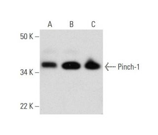
Pinch-1 Antibody (A-1): sc-393133
- Pinch-1 Antibody (A-1) is a mouse monoclonal IgG1 κ Pinch-1 antibody, cited in 5 publications, provided at 200 µg/ml
- specific for an epitope mapping between amino acids 132-169 within an internal region of Pinch-1 of human origin
- Pinch-1 Antibody (A-1) is recommended for detection of Pinch-1 of mouse, rat, human and avian origin by WB, IP, IF and ELISA; also reactive with additional species, including and canine, bovine, porcine and avian
- Anti-Pinch-1 Antibody (A-1) is available conjugated to agarose for IP; HRP for WB, IHC(P) and ELISA; and to either phycoerythrin or FITC for IF, IHC(P) and FCM
- also available conjugated to Alexa Fluor® 488, Alexa Fluor® 546, Alexa Fluor® 594 or Alexa Fluor® 647 for WB (RGB), IF, IHC(P) and FCM, and for use with RGB fluorescent imaging systems, such as iBright™ FL1000, FluorChem™, Typhoon, Azure and other comparable systems
- also available conjugated to Alexa Fluor® 680 or Alexa Fluor® 790 for WB (NIR), IF and FCM; for use with Near-Infrared (NIR) detection systems, such as LI-COR®Odyssey®, iBright™ FL1000, FluorChem™, Typhoon, Azure and other comparable systems
- m-IgG Fc BP-HRP, m-IgG1 BP-HRP and m-IgGκ BP-HRP are the preferred secondary detection reagents for Pinch-1 Antibody (A-1) for WB applications. These reagents are now offered in bundles with Pinch-1 Antibody (A-1) (see ordering information below).

QUICK LINKS
Pinch-1 Antibody (A-1) is a mouse monoclonal IgG1 kappa light chain antibody that detects Pinch-1 protein of mouse, rat, and human origin by western blotting (WB), immunoprecipitation (IP), immunofluorescence (IF), and enzyme-linked immunosorbent assay (ELISA). anti-Pinch-1 antibody (A-1) is available in both non-conjugated and various conjugated forms, including agarose, horseradish peroxidase (HRP), phycoerythrin (PE), fluorescein isothiocyanate (FITC), and multiple Alexa Fluor® conjugates. Pinch-1, also known as particularly interesting new Cys-His protein or NY-REN-48, plays a crucial role in cellular processes as a focal adhesion protein and a key component of the ILK-PINCH complex, which is integral to growth factor and integrin signaling pathways. This complex is essential for regulating cell differentiation, proliferation, and survival, as an effector of integrin and growth factor signaling. Pinch-1 is predominantly located in the cytoplasm and is expressed in a wide range of tissues, featuring a structure that includes five LIM domains, a nuclear localization signal, and a nuclear export signal, which are vital for cellular signaling. Additionally, PINCH-1/ILK complex activity is modulated by PINCH-2, which also interacts with ILK, highlighting these proteins′ importance in maintaining cellular homeostasis and responding to extracellular signals.
Alexa Fluor® is a trademark of Molecular Probes Inc., OR., USA
LI-COR® and Odyssey® are registered trademarks of LI-COR Biosciences
Pinch-1 Antibody (A-1) References:
- Characterization of PINCH-2, a new focal adhesion protein that regulates the PINCH-1-ILK interaction, cell spreading, and migration. | Zhang, Y., et al. 2002. J Biol Chem. 277: 38328-38. PMID: 12167643
- PINCH-1 is an obligate partner of integrin-linked kinase (ILK) functioning in cell shape modulation, motility, and survival. | Fukuda, T., et al. 2003. J Biol Chem. 278: 51324-33. PMID: 14551191
- Structure of an ultraweak protein-protein complex and its crucial role in regulation of cell morphology and motility. | Vaynberg, J., et al. 2005. Mol Cell. 17: 513-23. PMID: 15721255
- Formation and phosphorylation of the PINCH-1-integrin linked kinase-alpha-parvin complex are important for regulation of renal glomerular podocyte adhesion, architecture, and survival. | Yang, Y., et al. 2005. J Am Soc Nephrol. 16: 1966-76. PMID: 15872073
- Molecular dissection of PINCH-1 reveals a mechanism of coupling and uncoupling of cell shape modulation and survival. | Xu, Z., et al. 2005. J Biol Chem. 280: 27631-7. PMID: 15941716
- PINCH, N(i)ck and the ILK: network wiring at cell-matrix adhesions. | Wu, C. 2005. Trends Cell Biol. 15: 460-6. PMID: 16084094
- PINCH-1 expression during early avian embryogenesis: implications for neural crest and heart development. | Martinsen, BJ., et al. 2006. Dev Dyn. 235: 152-62. PMID: 16258920
- TGF-beta1 regulates the PINCH-1-integrin-linked kinase-alpha-parvin complex in glomerular cells. | Jung, KY., et al. 2007. J Am Soc Nephrol. 18: 66-73. PMID: 17167118
Ordering Information
| Product Name | Catalog # | UNIT | Price | Qty | FAVORITES | |
Pinch-1 Antibody (A-1) | sc-393133 | 200 µg/ml | $322.00 | |||
Pinch-1 Antibody (A-1): m-IgG Fc BP-HRP Bundle | sc-530365 | 200 µg Ab; 10 µg BP | $361.00 | |||
Pinch-1 Antibody (A-1): m-IgGκ BP-HRP Bundle | sc-523818 | 200 µg Ab, 40 µg BP | $361.00 | |||
Pinch-1 Antibody (A-1): m-IgG1 BP-HRP Bundle | sc-544016 | 200 µg Ab; 20 µg BP | $361.00 | |||
Pinch-1 Antibody (A-1) AC | sc-393133 AC | 500 µg/ml, 25% agarose | $424.00 | |||
Pinch-1 Antibody (A-1) HRP | sc-393133 HRP | 200 µg/ml | $322.00 | |||
Pinch-1 Antibody (A-1) FITC | sc-393133 FITC | 200 µg/ml | $336.00 | |||
Pinch-1 Antibody (A-1) PE | sc-393133 PE | 200 µg/ml | $349.00 | |||
Pinch-1 Antibody (A-1) Alexa Fluor® 488 | sc-393133 AF488 | 200 µg/ml | $364.00 | |||
Pinch-1 Antibody (A-1) Alexa Fluor® 546 | sc-393133 AF546 | 200 µg/ml | $364.00 | |||
Pinch-1 Antibody (A-1) Alexa Fluor® 594 | sc-393133 AF594 | 200 µg/ml | $364.00 | |||
Pinch-1 Antibody (A-1) Alexa Fluor® 647 | sc-393133 AF647 | 200 µg/ml | $364.00 | |||
Pinch-1 Antibody (A-1) Alexa Fluor® 680 | sc-393133 AF680 | 200 µg/ml | $364.00 | |||
Pinch-1 Antibody (A-1) Alexa Fluor® 790 | sc-393133 AF790 | 200 µg/ml | $364.00 | |||
Pinch-1 (A-1) Neutralizing Peptide | sc-393133 P | 100 µg/0.5 ml | $69.00 |