
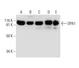


OPA1 Antibody (D-9): sc-393296
- OPA1 Antibody (D-9) is a mouse monoclonal IgG1 κ OPA1 antibody, cited in 62 publications, provided at 200 µg/ml
- raised against amino acids 647-780 mapping within an internal region of OPA1 of human origin
- OPA1 Antibody (D-9) is recommended for detection of OPA1 of mouse, rat and human origin by WB, IP, IF and ELISA; also reactive with additional species, including and equine, canine, bovine and porcine
- Anti-OPA1 Antibody (D-9) is available conjugated to agarose for IP; HRP for WB, IHC(P) and ELISA; and to either phycoerythrin or FITC for IF, IHC(P) and FCM
- also available conjugated to Alexa Fluor® 488, Alexa Fluor® 546, Alexa Fluor® 594 or Alexa Fluor® 647 for WB (RGB), IF, IHC(P) and FCM, and for use with RGB fluorescent imaging systems, such as iBright™ FL1000, FluorChem™, Typhoon, Azure and other comparable systems
- also available conjugated to Alexa Fluor® 680 or Alexa Fluor® 790 for WB (NIR), IF and FCM; for use with Near-Infrared (NIR) detection systems, such as LI-COR®Odyssey®, iBright™ FL1000, FluorChem™, Typhoon, Azure and other comparable systems
- m-IgG Fc BP-HRP and m-IgGκ BP-HRP are the preferred secondary detection reagents for OPA1 Antibody (D-9) for WB applications. These reagents are now offered in bundles with OPA1 Antibody (D-9) (see ordering information below).

QUICK LINKS
OPA1 Antibody (D-9) is a mouse monoclonal IgG1 kappa light chain antibody that detects OPA1 protein of mouse, rat, and human origin by western blotting (WB), immunoprecipitation (IP), immunofluorescence (IF), and enzyme-linked immunosorbent assay (ELISA). OPA1 Antibody (D-9) is available in both non-conjugated and various conjugated forms, including agarose, horseradish peroxidase (HRP), phycoerythrin (PE), fluorescein isothiocyanate (FITC), and multiple Alexa Fluor® conjugates. OPA1, encoded by the optic atrophy 1 gene located on chromosome 3q29, plays a crucial role in mitochondrial dynamics, particularly in regulating mitochondrial fusion and maintaining mitochondrial morphology. This function is vital for cellular energy production and overall cell health, helping ensure mitochondria can efficiently adapt to changes in energy demand and stress conditions. Defects in OPA1 are associated with optic atrophy type 1, leading to vision loss, and OPA1 is predominantly expressed in the retina, although also found in the brain, testis, heart, and skeletal muscles. OPA1′s ability to interact with other proteins involved in mitochondrial function further underscores OPA1′s importance in cellular metabolism and the pathophysiology of mitochondrial diseases.
Alexa Fluor® is a trademark of Molecular Probes Inc., OR., USA
LI-COR® and Odyssey® are registered trademarks of LI-COR Biosciences
OPA1 Antibody (D-9) References:
- Nuclear gene OPA1, encoding a mitochondrial dynamin-related protein, is mutated in dominant optic atrophy. | Delettre, C., et al. 2000. Nat Genet. 26: 207-10. PMID: 11017079
- Spectrum, frequency and penetrance of OPA1 mutations in dominant optic atrophy. | Toomes, C., et al. 2001. Hum Mol Genet. 10: 1369-78. PMID: 11440989
- Mutation spectrum and splicing variants in the OPA1 gene. | Delettre, C., et al. 2001. Hum Genet. 109: 584-91. PMID: 11810270
- Differential sublocalization of the dynamin-related protein OPA1 isoforms in mitochondria. | Satoh, M., et al. 2003. Biochem Biophys Res Commun. 300: 482-93. PMID: 12504110
- Roles of the mammalian mitochondrial fission and fusion mediators Fis1, Drp1, and Opa1 in apoptosis. | Lee, YJ., et al. 2004. Mol Biol Cell. 15: 5001-11. PMID: 15356267
- OPA1 requires mitofusin 1 to promote mitochondrial fusion. | Cipolat, S., et al. 2004. Proc Natl Acad Sci U S A. 101: 15927-32. PMID: 15509649
- OPA1, associated with autosomal dominant optic atrophy, is widely expressed in the human brain. | Bette, S., et al. 2005. Acta Neuropathol. 109: 393-9. PMID: 15700187
- Refinement of the dominant optic atrophy locus (OPA1) to a 1.4-cM interval on chromosome 3q28-3q29, within a 3-Mb YAC contig. | Jonasdottir, A., et al. 1997. Hum Genet. 99: 115-20. PMID: 9003507
Ordering Information
| Product Name | Catalog # | UNIT | Price | Qty | FAVORITES | |
OPA1 Antibody (D-9) | sc-393296 | 200 µg/ml | $322.00 | |||
OPA1 Antibody (D-9): m-IgG Fc BP-HRP Bundle | sc-530422 | 200 µg Ab; 10 µg BP | $361.00 | |||
OPA1 Antibody (D-9): m-IgGκ BP-HRP Bundle | sc-523893 | 200 µg Ab, 40 µg BP | $361.00 | |||
OPA1 Antibody (D-9) AC | sc-393296 AC | 500 µg/ml, 25% agarose | $424.00 | |||
OPA1 Antibody (D-9) HRP | sc-393296 HRP | 200 µg/ml | $322.00 | |||
OPA1 Antibody (D-9) FITC | sc-393296 FITC | 200 µg/ml | $336.00 | |||
OPA1 Antibody (D-9) PE | sc-393296 PE | 200 µg/ml | $349.00 | |||
OPA1 Antibody (D-9) Alexa Fluor® 488 | sc-393296 AF488 | 200 µg/ml | $364.00 | |||
OPA1 Antibody (D-9) Alexa Fluor® 546 | sc-393296 AF546 | 200 µg/ml | $364.00 | |||
OPA1 Antibody (D-9) Alexa Fluor® 594 | sc-393296 AF594 | 200 µg/ml | $364.00 | |||
OPA1 Antibody (D-9) Alexa Fluor® 647 | sc-393296 AF647 | 200 µg/ml | $364.00 | |||
OPA1 Antibody (D-9) Alexa Fluor® 680 | sc-393296 AF680 | 200 µg/ml | $364.00 | |||
OPA1 Antibody (D-9) Alexa Fluor® 790 | sc-393296 AF790 | 200 µg/ml | $364.00 |