")
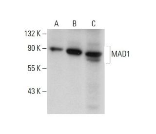
MAD1 Antibody (D-1): sc-166312
- MAD1 Antibody (D-1) is a mouse monoclonal IgG2b provided at 200 µg/ml
- raised against amino acids 491-718 mapping at the C-terminus of MAD1 of human origin
- recommended for detection of MAD1 of mouse, rat and human origin by WB, IP, IF, IHC(P) and ELISA
- m-IgG Fc BP-HRP, m-IgG2b BP-HRP and m-IgGκ BP-HRP are the preferred secondary detection reagents for MAD1 Antibody (D-1) for WB and IHC(P) applications. These reagents are now offered in bundles with MAD1 Antibody (D-1) (see ordering information below).

QUICK LINKS
MAD1 Antibody (D-1) is a mouse monoclonal IgG2b antibody that detects MAD1 in mouse, rat, and human samples through various applications including western blotting (WB), immunoprecipitation (IP), immunofluorescence (IF), immunohistochemistry with paraffin-embedded sections (IHCP), and enzyme-linked immunosorbent assay (ELISA). MAD1 plays a critical role in cell cycle regulation by ensuring proper chromosome alignment at the mitotic spindle assembly checkpoint, preventing premature anaphase onset. This function is vital for maintaining genomic stability, as errors in chromosome segregation can lead to aneuploidy and contribute to cancer development. MAD1 is primarily localized in the nucleus, where MAD1 anchors MAD2L1 to the nuclear periphery, but during mitosis, MAD1 dynamically relocates to the centrosome, spindle midzone, and midbody, highlighting MAD1′s importance in the spatial regulation of mitotic processes. Additionally, MAD1 can form homo- and heterodimers with MAD2, contributing to the formation of the tetrameric MAD1L1-MAD2L1 core complex, which is essential for the checkpoint′s functionality. MAD1 activity is further modulated by post-translational modifications, including hyperphosphorylation in response to spindle damage, underscoring MAD1′s role in the cellular response to mitotic stress. Defects in the MAD1 gene, MAD1L1, are implicated in various cancers, making MAD1 (D-1) antibody a valuable tool for studying cell cycle dynamics and cancer biology.
Ordering Information
| Product Name | Catalog # | UNIT | Price | Qty | FAVORITES | |
MAD1 Antibody (D-1) | sc-166312 | 200 µg/ml | $322.00 | |||
MAD1 Antibody (D-1): m-IgG Fc BP-HRP Bundle | sc-537473 | 200 µg Ab; 10 µg BP | $361.00 | |||
MAD1 Antibody (D-1): m-IgGκ BP-HRP Bundle | sc-534751 | 200 µg Ab; 40 µg BP | $361.00 | |||
MAD1 Antibody (D-1): m-IgG2b BP-HRP Bundle | sc-549878 | 200 µg Ab; 10 µg BP | $361.00 |