")
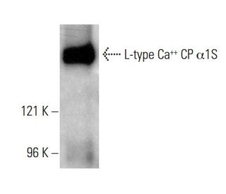
: sc-21781. Western blot analysis of L-type Ca<sup>++</sup> CP α1S expression in rat skeletal muscle tissue extract.")
L-type Ca++ CP α1S Antibody (IIC12D4): sc-21781
- L-type Ca++ CP α1S Antibody (IIC12D4) is a mouse monoclonal IgG1 κ L-type Ca++ CP α1S antibody, cited in 2 publications, provided at 200 µg/ml
- raised against rabbit skeletal muscle triads
- recommended for detection of 170 kDa, L-type calcium channel α1S of mouse, rat, human and rabbit origin by WB, IP and IF
- m-IgG Fc BP-HRP, m-IgG1 BP-HRP and m-IgGκ BP-HRP are the preferred secondary detection reagents for L-type Ca++ CP α1S Antibody (IIC12D4) for WB applications. These reagents are now offered in bundles with L-type Ca++ CP α1S Antibody (IIC12D4) (see ordering information below).

QUICK LINKS
SEE ALSO...
L-type Ca++ CP α1S Antibody (IIC12D4) is a mouse monoclonal IgG1 kappa light chain antibody that detects the L-type Ca++ CP alpha 1S protein of mouse, rat, human, and rabbit origin by western blotting (WB), immunoprecipitation (IP), and immunofluorescence (IF). Anti-L-type Ca++ CP alpha 1S antibody (IIC12D4) is available as the non-conjugated form. The L-type calcium channel plays a crucial role in regulating calcium ion entry into excitable cells, which is essential for various physiological processes such as muscle contraction, hormone secretion, and neurotransmitter release. These channels are composed of a complex structure that includes an alpha-1 subunit, which forms the channel pore, along with auxiliary subunits that modulate channel activity and pharmacological properties. The alpha-1S subunit is vital for excitation-contraction coupling in skeletal muscle, as this subunit directly influences calcium release from the sarcoplasmic reticulum, thereby initiating muscle contraction. Additionally, L-type calcium channels can interact with various signaling molecules, including G protein-coupled receptors, which enhances regulatory capacity and specificity in cellular signaling pathways. This intricate interplay underscores the importance of L-type Ca++ CP alpha 1S protein in maintaining cellular function and responding to physiological demands.
Alexa Fluor® is a trademark of Molecular Probes Inc., OR., USA
LI-COR® and Odyssey® are registered trademarks of LI-COR Biosciences
L-type Ca++ CP α1S Antibody (IIC12D4) References:
- Cav1.1 controls frequency-dependent events regulating adult skeletal muscle plasticity. | Jorquera, G., et al. 2013. J Cell Sci. 126: 1189-98. PMID: 23321639
- Apparent lack of physical or functional interaction between CaV1.1 and its distal C terminus. | Ohrtman, JD., et al. 2015. J Gen Physiol. 145: 303-14. PMID: 25779869
- Structure of the voltage-gated calcium channel Cav1.1 complex. | Wu, J., et al. 2015. Science. 350: aad2395. PMID: 26680202
- Progressive impairment of CaV1.1 function in the skeletal muscle of mice expressing a mutant type 1 Cu/Zn superoxide dismutase (G93A) linked to amyotrophic lateral sclerosis. | Beqollari, D., et al. 2016. Skelet Muscle. 6: 24. PMID: 27340545
- Distinct transcriptomic changes in E14.5 mouse skeletal muscle lacking RYR1 or Cav1.1 converge at E18.5. | Filipova, D., et al. 2018. PLoS One. 13: e0194428. PMID: 29543863
- Gating pore currents occur in CaV1.1 domain III mutants associated with HypoPP. | Wu, F., et al. 2021. J Gen Physiol. 153: PMID: 34463712
- The distinct role of the four voltage sensors of the skeletal CaV1.1 channel in voltage-dependent activation. | Savalli, N., et al. 2021. J Gen Physiol. 153: PMID: 34546289
- Distinct roles for CaV1.1's voltage-sensing domains. | Short, B. 2021. J Gen Physiol. 153: PMID: 34623381
- Pannexin-1 and CaV1.1 show reciprocal interaction during excitation-contraction and excitation-transcription coupling in skeletal muscle. | Jaque-Fernández, F., et al. 2021. J Gen Physiol. 153: PMID: 34636893
- Calcium current modulation by the γ1 subunit depends on alternative splicing of CaV1.1. | El Ghaleb, Y., et al. 2022. J Gen Physiol. 154: PMID: 35349630
Ordering Information
| Product Name | Catalog # | UNIT | Price | Qty | FAVORITES | |
L-type Ca++ CP α1S Antibody (IIC12D4) | sc-21781 | 200 µg/ml | $322.00 | |||
L-type Ca++ CP α1S Antibody (IIC12D4): m-IgG Fc BP-HRP Bundle | sc-536672 | 200 µg Ab; 10 µg BP | $361.00 | |||
L-type Ca++ CP α1S Antibody (IIC12D4): m-IgGκ BP-HRP Bundle | sc-533831 | 200 µg Ab; 40 µg BP | $361.00 | |||
L-type Ca++ CP α1S Antibody (IIC12D4): m-IgG1 BP-HRP Bundle | sc-544754 | 200 µg Ab; 20 µg BP | $361.00 |