")
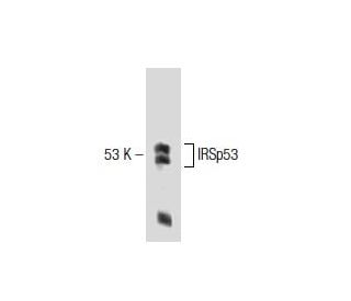
IRSp53 Antibody (46): sc-136470
- IRSp53 Antibody (46) is a mouse monoclonal IgG2a κ IRSp53 antibody, cited in 3 publications, provided at 200 µg/ml
- raised against amino acids 200-322 of IRSp53 of human origin
- recommended for detection of IRSp53 of mouse, rat and human origin by WB and IP; also reactive with additional species, including canine
- available conjugated to agarose for IP; and to HRP for WB, IHC(P) and ELISA
- m-IgG Fc BP-HRP, m-IgG2a BP-HRP and m-IgGκ BP-HRP are the preferred secondary detection reagents for IRSp53 Antibody (46) for WB applications. These reagents are now offered in bundles with IRSp53 Antibody (46) (see ordering information below).

QUICK LINKS
SEE ALSO...
IRSp53 Antibody (46) is a mouse monoclonal IgG2a kappa light chain antibody that detects IRSp53 protein of mouse, rat, and human origin by western blotting (WB) and immunoprecipitation (IP). Anti-IRSp53 antibody (46) is available in both non-conjugated and various conjugated forms, including agarose and horseradish peroxidase (HRP). The insulin receptor tyrosine kinase substrate p53 (IRSp53) is a crucial scaffolding protein that plays a significant role in regulating the actin cytoskeleton, particularly in mediating filopodia formation under the influence of Rho-family GTPases. IRSp53 is predominantly located in the cytoplasm, where IRSp53 serves as a vital link between small membrane-bound G-proteins and cytoplasmic effector proteins, facilitating cellular signaling pathways essential for cell motility and morphology. Structurally, IRSp53 features a central SH3 domain that interacts with proline-rich regions of various actin regulators, alongside a conserved N-terminal IRSp53/MIM homology domain (IMD) that exhibits F-actin-bundling activity. IRSp53 interacts with several key proteins, including atrophin-1, which is associated with dentatorubral-pallidoluysian atrophy, as well as ENAH, BAI-1, Eps8, and the Shank family of proteins (Shank 1, Shank 2, Shank 3), along with WAVE1, WAVE2, Tiam1, and Dia 1. These interactions underscore IRSp53′s importance in maintaining cellular architecture and signaling, making IRSp53 monoclonal antibody (46) an invaluable tool for researchers studying cytoskeletal dynamics and related pathologies.
Alexa Fluor® is a trademark of Molecular Probes Inc., OR., USA
LI-COR® and Odyssey® are registered trademarks of LI-COR Biosciences
IRSp53 Antibody (46) References:
- Dentatorubral-pallidoluysian atrophy protein interacts through a proline-rich region near polyglutamine with the SH3 domain of an insulin receptor tyrosine kinase substrate. | Okamura-Oho, Y., et al. 1999. Hum Mol Genet. 8: 947-57. PMID: 10332026
- The insulin receptor substrate IRSp53 links postsynaptic shank1 to the small G-protein cdc42. | Soltau, M., et al. 2002. Mol Cell Neurosci. 21: 575-83. PMID: 12504591
- Genomic structure and alternative splicing of the insulin receptor tyrosine kinase substrate of 53-kDa protein. | Miyahara, A., et al. 2003. J Hum Genet. 48: 410-414. PMID: 12884081
- IRSp53/Eps8 complex is important for positive regulation of Rac and cancer cell motility/invasiveness. | Funato, Y., et al. 2004. Cancer Res. 64: 5237-44. PMID: 15289329
- Structural basis of filopodia formation induced by the IRSp53/MIM homology domain of human IRSp53. | Millard, TH., et al. 2005. EMBO J. 24: 240-50. PMID: 15635447
- Regulation of dendritic spine morphogenesis by insulin receptor substrate 53, a downstream effector of Rac1 and Cdc42 small GTPases. | Choi, J., et al. 2005. J Neurosci. 25: 869-79. PMID: 15673667
- NMDA receptor-dependent synaptic translocation of insulin receptor substrate p53 via protein kinase C signaling. | Hori, K., et al. 2005. J Neurosci. 25: 2670-81. PMID: 15758177
- Tiam1-IRSp53 complex formation directs specificity of rac-mediated actin cytoskeleton regulation. | Connolly, BA., et al. 2005. Mol Cell Biol. 25: 4602-14. PMID: 15899863
- Novel brain 14-3-3 interacting proteins involved in neurodegenerative disease. | Mackie, S. and Aitken, A. 2005. FEBS J. 272: 4202-10. PMID: 16098201
- IRSp53/BAIAP2 in dendritic spine development, NMDA receptor regulation, and psychiatric disorders. | Kang, J., et al. 2016. Neuropharmacology. 100: 27-39. PMID: 26275848
- IRSp53 promotes postsynaptic density formation and actin filament bundling. | Feng, Z., et al. 2022. J Cell Biol. 221: PMID: 35819332
- Tunnelling nanotube formation is driven by Eps8/IRSp53-dependent linear actin polymerization. | Henderson, JM., et al. 2023. EMBO J. 42: e113761. PMID: 38009333
Ordering Information
| Product Name | Catalog # | UNIT | Price | Qty | FAVORITES | |
IRSp53 Antibody (46) | sc-136470 | 200 µg/ml | $322.00 | |||
IRSp53 Antibody (46): m-IgG Fc BP-HRP Bundle | sc-528828 | 200 µg Ab; 10 µg BP | $361.00 | |||
IRSp53 Antibody (46): m-IgGκ BP-HRP Bundle | sc-521373 | 200 µg Ab, 40 µg BP | $361.00 | |||
IRSp53 Antibody (46): m-IgG2a BP-HRP Bundle | sc-547179 | 200 µg Ab; 10 µg BP | $361.00 | |||
IRSp53 Antibody (46) AC | sc-136470 AC | 500 µg/ml, 25% agarose | $424.00 | |||
IRSp53 Antibody (46) HRP | sc-136470 HRP | 200 µg/ml | $322.00 |