Date published: 2026-4-25

1-800-457-3801

SCBT Portrait Logo
Seach Input

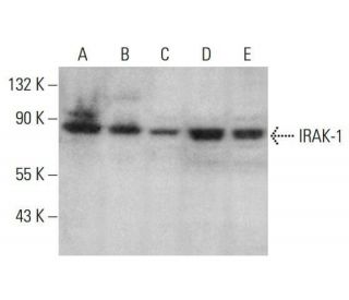
IRAK-1 Antibody (C-2): sc-5287

4.7(6)
Write a reviewAsk a question

Datasheets
  • IRAK-1 Antibody (C-2) is a mouse monoclonal IgG2a κ, cited in 8 publications, provided at 200 µg/ml
  • raised against amino acids 440-712 of IRAK-1 of human origin
  • recommended for detection of IRAK-1 of human origin by WB, IP, IF, IHC(P) and ELISA
  • See IRAK-1 (F-4): sc-5288 for IRAK-1 antibody conjugates, including AC, HRP, FITC, PE, Alexa Fluor® 488, 594, 647, 680 and 790.
  • m-IgG Fc BP-HRP, m-IgG2a BP-HRP and m-IgGκ BP-HRP are the preferred secondary detection reagents for IRAK-1 Antibody (C-2) for WB and IHC(P) applications. These reagents are now offered in bundles with IRAK-1 Antibody (C-2) (see ordering information below).
Gene Editing Promo Banner

QUICK LINKS

SEE ALSO...

IRAK-1 Antibody (C-2) is a mouse monoclonal IgG2a antibody that detects IRAK-1 in human samples through various applications including western blotting (WB), immunoprecipitation (IP), immunofluorescence (IF), immunohistochemistry with paraffin embedded sections (IHCP), and enzyme-linked immunosorbent assay (ELISA). IRAK-1, or IL-1 receptor-associated kinase 1, plays a crucial role in the signaling pathways activated by interleukin-1 (IL-1), particularly in the activation of the NF-kB pathway, which is essential for regulating immune responses and inflammation. Located primarily in the cytoplasm, IRAK-1 is recruited to the IL-1 receptor complex upon ligand binding, where IRAK-1 undergoes phosphorylation and subsequently activates downstream signaling cascades. This process is vital for the propagation of inflammatory signals, and dysregulation of IRAK-1 has been implicated in various inflammatory diseases and conditions, making IRAK-1 a significant target for therapeutic intervention. The ability of IRAK-1 to interact with other proteins, such as TRAF6 and MyD88, further underscores IRAK-1′s importance in the immune response, as these interactions facilitate the assembly of signaling complexes that lead to the activation of transcription factors like NF-kB. IRAK-1 (C-2) antibody is an invaluable tool for researchers studying the intricate mechanisms of immune signaling and inflammation.

For Research Use Only. Not Intended for Diagnostic or Therapeutic Use.

Ordering Information

Product NameCatalog #UNITPriceQtyFAVORITES

IRAK-1 Antibody (C-2)

sc-5287
200 µg/ml
$322.00

IRAK-1 Antibody (C-2): m-IgG Fc BP-HRP Bundle

sc-536547
200 µg Ab; 10 µg BP
$361.00

IRAK-1 Antibody (C-2): m-IgGκ BP-HRP Bundle

sc-533698
200 µg Ab; 40 µg BP
$361.00

IRAK-1 Antibody (C-2): m-IgG2a BP-HRP Bundle

sc-547834
200 µg Ab; 10 µg BP
$361.00

I am using IRAK-1 (C-2): sc-5287 monoclonal antibody to stain paraffin embedded tissue. Do you recommend antigen retrieval when using this antibody?

Asked by: TinTin
We recommend performing antigen retrieval with sodium citrate buffer (pH 6) and heat. The full protocol can be found here: https://www.scbt.com/scbt/resources/protocols/immunoperoxidase-staining
Answered by: Technical Support
Date published: 2017-02-24
  • y_2026, m_4, d_24, h_10CST
  • bvseo_bulk, prod_bvqa, vn_bulk_3.0.43
  • cp_1, bvpage1
  • co_hasquestionsanswers, tq_1
  • loc_en_US, sid_5287, prod, sort_[SortEntry(order=LAST_APPROVED_ANSWER_SUBMISSION_TIME, direction=DESCENDING)]
  • clientName_scbt
  • bvseo_sdk, java_sdk, bvseo-4.0.0
  • CLOUD, getContent, 107ms
  • QUESTIONS, PRODUCT
Rated 5 out of 5 by from Nice antibody for IRAK1 detection in HeLa cellsWe used this antibody for expression detection of IRAK1 by western blot. We got a very nice signal around the correct molecular weight in HeLa cells.
Date published: 2017-02-09
Rated 5 out of 5 by from Superb immunoperoxidase cytoplasmic and membraneSuperb immunoperoxidase cytoplasmic and membrane staining in formalin fixed, paraffin-embedded human breast tissue. -SCBT QC
Date published: 2015-05-09
Rated 5 out of 5 by from Great band detected in WB using T24 wholeGreat band detected in WB using T24 whole cell lysate. -SCBT QC
Date published: 2015-04-20
Rated 4 out of 5 by from Various publishable data in WB using humanVarious publishable data in WB using human Mono Mac 6 cells, Monocyte, HPC-4, DeTa, HCV29, A549, Jurkat, Raji, HAE, breast carcinoma, pancreatic adenocarcinoma cells and normal bronchial epithelium cells -SCBT Publication Review
Date published: 2015-04-04
Rated 5 out of 5 by from Strong band seen in WB using KStrong band seen in WB using K-562 nuclear extract. -SCBT QC
Date published: 2015-01-13
Rated 4 out of 5 by from Publishable data in WB using mouse transfectedPublishable data in WB using mouse transfected peritoneal macrophages -SCBT Publication Review
Date published: 2015-01-11
  • y_2026, m_4, d_24, h_10
  • bvseo_bulk, prod_bvrr, vn_bulk_3.0.43
  • cp_1, bvpage1
  • co_hasreviews, tv_0, tr_6
  • loc_en_US, sid_5287, prod, sort_[SortEntry(order=SUBMISSION_TIME, direction=DESCENDING)]
  • clientName_scbt
  • bvseo_sdk, java_sdk, bvseo-4.0.0
  • CLOUD, getReviews, 16ms
  • REVIEWS, PRODUCT
IRAK-1 Antibody (C-2) is rated 4.7 out of 5 by 6.
  • y_2026, m_4, d_24, h_10
  • bvseo_bulk, prod_bvrr, vn_bulk_3.0.43
  • cp_1, bvpage1
  • co_hasreviews, tv_0, tr_6
  • loc_en_US, sid_5287, prod, sort_[SortEntry(order=SUBMISSION_TIME, direction=DESCENDING)]
  • clientName_scbt
  • bvseo_sdk, java_sdk, bvseo-4.0.0
  • CLOUD, getAggregateRating, 103ms
  • REVIEWS, PRODUCT