")
Id1 Antibody (B-8): sc-133104
- Id1 Antibody (B-8) is a mouse monoclonal IgG2a κ Id1 antibody, cited in 43 publications, provided at 200 µg/ml
- specific for an epitope mapping between amino acids 118-159 at the C-terminus of Id1 of mouse origin
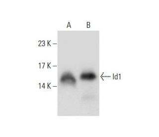
- Id1 Antibody (B-8) is recommended for detection of Id1 of mouse, rat and human origin by WB, IP, IF, IHC(P) and ELISA; also reactive with additional species, including canine
- Anti-Id1 Antibody (B-8) is available conjugated to agarose for IP; HRP for WB, IHC(P) and ELISA; and to either phycoerythrin or FITC for IF, IHC(P) and FCM
- also available conjugated to Alexa Fluor® 488, Alexa Fluor® 546, Alexa Fluor® 594 or Alexa Fluor® 647 for WB (RGB), IF, IHC(P) and FCM, and for use with RGB fluorescent imaging systems, such as iBright™ FL1000, FluorChem™, Typhoon, Azure and other comparable systems
- also available conjugated to Alexa Fluor® 680 or Alexa Fluor® 790 for WB (NIR), IF and FCM; for use with Near-Infrared (NIR) detection systems, such as LI-COR®Odyssey®, iBright™ FL1000, FluorChem™, Typhoon, Azure and other comparable systems
- TransCruz reagent for Gel Supershift and ChIP applications (sc-133104 X, 200 µg/0.1 ml)
- m-IgG Fc BP-HRP and m-IgGκ BP-HRP are the preferred secondary detection reagents for Id1 Antibody (B-8) for WB and IHC(P) applications. These reagents are now offered in bundles with Id1 Antibody (B-8) (see ordering information below).

QUICK LINKS
Id1 Antibody (B-8) is a mouse monoclonal IgG2a kappa light chain antibody engineered to target the C-terminal epitope (amino acids 118-159) of the mouse Id1 protein. Inhibitor of DNA binding 1 (Id1), also called Idb1, bHLHb24, and D2Wsu140e, is a pivotal member of the Inhibitor of DNA binding (Id) family of basic helix-loop-helix (bHLH) proteins, and plays a critical role in regulating various cellular processes including differentiation, proliferation, and angiogenesis. Located primarily in the nucleus, Id1 functions by dimerizing with class A and B HLH proteins, thereby inhibiting their ability to bind DNA and negatively regulating their transcriptional activity. This nuclear localization is essential as Id1 controls the expression of genes involved in muscle-specific differentiation by preventing transcription factors like E12 and Myo D from activating myogenic genes. Under conditions that promote muscle cell differentiation, nuclear levels of Id1 decrease, facilitating the formation of active heterodimers between E12 or E47 and Myo D or myogenin, which then drive the differentiation process forward. Additionally, Id1 is integral to cell cycle regulation, with expression being tightly controlled by growth factor signals. Reduction of Id1 mRNA levels has been shown to delay the reentry of arrested cells into the cell cycle following growth factor stimulation, highlighting Id1′s importance in cell proliferation and cancer research. Anti-Id1 antibody (B-8) exhibits broad species reactivity, effectively recognizing Id1 proteins from mouse, rat, and human origins, as well as additional species including canine. Id1 (B-8) antibody is versatile in multiple applications such as western blotting (WB), immunoprecipitation (IP), immunofluorescence (IF), immunohistochemistry with paraffin-embedded sections (IHCP), and enzyme-linked immunosorbent assay (ELISA), making Id1 (B-8) monoclonal antibody an invaluable tool for researchers investigating the intricate roles of Id1 in various biological contexts. By providing detailed insights into Id1′s cellular localization and function, anti-Id1 antibody (B-8) supports advancements in understanding muscle differentiation, cell cycle control, and the molecular mechanisms underlying a range of diseases.
Alexa Fluor® is a trademark of Molecular Probes Inc., OR., USA
LI-COR® and Odyssey® are registered trademarks of LI-COR Biosciences
Id1 Antibody (B-8) References:
- Id proteins Id1 and Id2 selectively inhibit DNA binding by one class of helix-loop-helix proteins. | Sun, XH., et al. 1991. Mol Cell Biol. 11: 5603-11. PMID: 1922066
- An Id-related helix-loop-helix protein encoded by a growth factor-inducible gene. | Christy, BA., et al. 1991. Proc Natl Acad Sci U S A. 88: 1815-9. PMID: 2000388
- The protein Id: a negative regulator of helix-loop-helix DNA binding proteins. | Benezra, R., et al. 1990. Cell. 61: 49-59. PMID: 2156629
- Id1 expression in kidney endothelial cells protects against diabetes-induced microvascular injury. | Sharma, S. and Plotkin, M. 2020. FEBS Open Bio. 10: 1447-1462. PMID: 31957231
- ID1 confers cancer cell chemoresistance through STAT3/ATF6-mediated induction of autophagy. | Meng, J., et al. 2020. Cell Death Dis. 11: 137. PMID: 32080166
- ID1 mediates resistance to osimertinib in EGFR T790M-positive non-small cell lung cancer through epithelial-mesenchymal transition. | Liu, K., et al. 2021. BMC Pulm Med. 21: 163. PMID: 33992097
- Therapeutic targeting of prenatal pontine ID1 signaling in diffuse midline glioma. | Messinger, D., et al. 2023. Neuro Oncol. 25: 54-67. PMID: 35605606
- Exopolysaccharide ID1 Improves Post-Warming Outcomes after Vitrification of In Vitro-Produced Bovine Embryos. | Ordóñez-León, EA., et al. 2022. Int J Mol Sci. 23: PMID: 35806071
- HLH forced dimers: tethering MyoD to E47 generates a dominant positive myogenic factor insulated from negative regulation by Id. | Neuhold, LA. and Wold, B. 1993. Cell. 74: 1033-42. PMID: 7691411
- The expression pattern of Id4, a novel dominant negative helix-loop-helix protein, is distinct from Id1, Id2 and Id3. | Riechmann, V., et al. 1994. Nucleic Acids Res. 22: 749-55. PMID: 8139914
- Id proteins control growth induction in mammalian cells. | Barone, MV., et al. 1994. Proc Natl Acad Sci U S A. 91: 4985-8. PMID: 8197168
- Id-related genes encoding helix-loop-helix proteins are required for G1 progression and are repressed in senescent human fibroblasts. | Hara, E., et al. 1994. J Biol Chem. 269: 2139-45. PMID: 8294468
Ordering Information
| Product Name | Catalog # | UNIT | Price | Qty | FAVORITES | |
Id1 Antibody (B-8) | sc-133104 | 200 µg/ml | $322.00 | |||
Id1 Antibody (B-8): m-IgG Fc BP-HRP Bundle | sc-528751 | 200 µg Ab; 10 µg BP | $361.00 | |||
Id1 Antibody (B-8): m-IgGκ BP-HRP Bundle | sc-521236 | 200 µg Ab, 40 µg BP | $361.00 | |||
Id1 Antibody (B-8) X | sc-133104 X | 200 µg/0.1 ml | $322.00 | |||
Id1 Antibody (B-8) AC | sc-133104 AC | 500 µg/ml, 25% agarose | $424.00 | |||
Id1 Antibody (B-8) HRP | sc-133104 HRP | 200 µg/ml | $322.00 | |||
Id1 Antibody (B-8) FITC | sc-133104 FITC | 200 µg/ml | $336.00 | |||
Id1 Antibody (B-8) PE | sc-133104 PE | 200 µg/ml | $349.00 | |||
Id1 Antibody (B-8) Alexa Fluor® 488 | sc-133104 AF488 | 200 µg/ml | $364.00 | |||
Id1 Antibody (B-8) Alexa Fluor® 546 | sc-133104 AF546 | 200 µg/ml | $364.00 | |||
Id1 Antibody (B-8) Alexa Fluor® 594 | sc-133104 AF594 | 200 µg/ml | $364.00 | |||
Id1 Antibody (B-8) Alexa Fluor® 647 | sc-133104 AF647 | 200 µg/ml | $364.00 | |||
Id1 Antibody (B-8) Alexa Fluor® 680 | sc-133104 AF680 | 200 µg/ml | $364.00 | |||
Id1 Antibody (B-8) Alexa Fluor® 790 | sc-133104 AF790 | 200 µg/ml | $364.00 | |||
Id1 (B-8) Neutralizing Peptide | sc-133104 P | 100 µg/0.5 ml | $69.00 |