")
DR3 Antibody (B-8): sc-374203
- DR3 Antibody (B-8) is a mouse monoclonal IgG2b κ DR3 antibody, cited in 3 publications, provided at 200 µg/ml
- specific for an epitope mapping between amino acids 387-416 at the C-terminus of DR3 of human origin
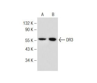
- DR3 Antibody (B-8) is recommended for detection of DR3 of mouse, rat and human origin by WB, IP, IF, IHC(P) and ELISA
- Anti-DR3 Antibody (B-8) is available conjugated to agarose for IP; HRP for WB, IHC(P) and ELISA; and to either phycoerythrin or FITC for IF, IHC(P) and FCM
- also available conjugated to Alexa Fluor® 488, Alexa Fluor® 546, Alexa Fluor® 594 or Alexa Fluor® 647 for WB (RGB), IF, IHC(P) and FCM, and for use with RGB fluorescent imaging systems, such as iBright™ FL1000, FluorChem™, Typhoon, Azure and other comparable systems
- also available conjugated to Alexa Fluor® 680 or Alexa Fluor® 790 for WB (NIR), IF and FCM; for use with Near-Infrared (NIR) detection systems, such as LI-COR®Odyssey®, iBright™ FL1000, FluorChem™, Typhoon, Azure and other comparable systems
- m-IgG2b BP-HRP and m-IgGκ BP-HRP are the preferred secondary detection reagents for DR3 Antibody (B-8) for WB and IHC(P) applications. These reagents are now offered in bundles with DR3 Antibody (B-8) (see ordering information below).

QUICK LINKS
DR3 Antibody (B-8) is a mouse monoclonal IgG2b kappa light chain antibody that detects DR3 protein of mouse, rat, and human origin by western blotting (WB), immunoprecipitation (IP), immunofluorescence (IF), immunohistochemistry, and enzyme-linked immunosorbent assay (ELISA). anti-DR3 antibody (B-8) is available in both non-conjugated and various conjugated forms, including agarose, horseradish peroxidase (HRP), phycoerythrin (PE), fluorescein isothiocyanate (FITC), and multiple Alexa Fluor® conjugates. DR3, also known as TNFRSF25, plays a crucial role in the immune system by mediating apoptosis and activating the NFκB signaling pathway, which is essential for regulating immune responses and maintaining homeostasis. Located primarily in thymocytes and lymphocytes, DR3 is a member of the tumor necrosis factor receptor superfamily and is characterized by its unique death domain that triggers apoptotic signaling. This specific localization is vital as DR3 effectively participates in the development and activation of T cells, thereby influencing adaptive immunity. DR3′s ability to induce apoptosis in certain immune cells highlights its importance in preventing autoimmune responses and maintaining immune tolerance. With versatile applications and critical role in immune regulation, DR3 (B-8) monoclonal antibody serves as an invaluable tool for researchers studying immune system dynamics and related pathologies.
Alexa Fluor® is a trademark of Molecular Probes Inc., OR., USA
LI-COR® and Odyssey® are registered trademarks of LI-COR Biosciences
DR3 Antibody (B-8) References:
- The role of TL1A and DR3 in autoimmune and inflammatory diseases. | Aiba, Y. and Nakamura, M. 2013. Mediators Inflamm. 2013: 258164. PMID: 24453414
- HLA-DR3 antigen in the resistance to idiopathic dilated cardiomyopathy. | Jin, B., et al. 2016. Braz J Med Biol Res. 49: e5131. PMID: 27007655
- The TL1A-DR3 Axis in Asthma: Membrane-Bound and Secreted TL1A Co-Determined the Development of Airway Remodeling. | Zhang, J., et al. 2022. Allergy Asthma Immunol Res. 14: 233-253. PMID: 35255540
- The Fas death factor. | Nagata, S. and Golstein, P. 1995. Science. 267: 1449-56. PMID: 7533326
- The TNF receptor superfamily of cellular and viral proteins: activation, costimulation, and death. | Smith, CA., et al. 1994. Cell. 76: 959-62. PMID: 8137429
- A novel domain within the 55 kd TNF receptor signals cell death. | Tartaglia, LA., et al. 1993. Cell. 74: 845-53. PMID: 8397073
- Apoptosis mediated by the TNF-related cytokine and receptor families. | Ware, CF., et al. 1996. J Cell Biochem. 60: 47-55. PMID: 8825415
- Signal transduction by DR3, a death domain-containing receptor related to TNFR-1 and CD95. | Chinnaiyan, AM., et al. 1996. Science. 274: 990-2. PMID: 8875942
- A death-domain-containing receptor that mediates apoptosis. | Kitson, J., et al. 1996. Nature. 384: 372-5. PMID: 8934525
- Apo-3, a new member of the tumor necrosis factor receptor family, contains a death domain and activates apoptosis and NF-kappa B. | Marsters, SA., et al. 1996. Curr Biol. 6: 1669-76. PMID: 8994832
- TRAMP, a novel apoptosis-mediating receptor with sequence homology to tumor necrosis factor receptor 1 and Fas(Apo-1/CD95). | Bodmer, JL., et al. 1997. Immunity. 6: 79-88. PMID: 9052839
- A new death receptor 3 isoform: expression in human lymphoid cell lines and non-Hodgkin's lymphomas. | Warzocha, K., et al. 1998. Biochem Biophys Res Commun. 242: 376-9. PMID: 9446802
Ordering Information
| Product Name | Catalog # | UNIT | Price | Qty | FAVORITES | |
DR3 Antibody (B-8) | sc-374203 | 200 µg/ml | $322.00 | |||
DR3 Antibody (B-8): m-IgGκ BP-HRP Bundle | sc-522659 | 200 µg Ab, 40 µg BP | $361.00 | |||
DR3 Antibody (B-8): m-IgG2b BP-HRP Bundle | sc-549052 | 200 µg Ab; 10 µg BP | $361.00 | |||
DR3 Antibody (B-8) AC | sc-374203 AC | 500 µg/ml, 25% agarose | $424.00 | |||
DR3 Antibody (B-8) HRP | sc-374203 HRP | 200 µg/ml | $322.00 | |||
DR3 Antibody (B-8) FITC | sc-374203 FITC | 200 µg/ml | $336.00 | |||
DR3 Antibody (B-8) PE | sc-374203 PE | 200 µg/ml | $349.00 | |||
DR3 Antibody (B-8) Alexa Fluor® 488 | sc-374203 AF488 | 200 µg/ml | $364.00 | |||
DR3 Antibody (B-8) Alexa Fluor® 546 | sc-374203 AF546 | 200 µg/ml | $364.00 | |||
DR3 Antibody (B-8) Alexa Fluor® 594 | sc-374203 AF594 | 200 µg/ml | $364.00 | |||
DR3 Antibody (B-8) Alexa Fluor® 647 | sc-374203 AF647 | 200 µg/ml | $364.00 | |||
DR3 Antibody (B-8) Alexa Fluor® 680 | sc-374203 AF680 | 200 µg/ml | $364.00 | |||
DR3 Antibody (B-8) Alexa Fluor® 790 | sc-374203 AF790 | 200 µg/ml | $364.00 | |||
DR3 (B-8) Neutralizing Peptide | sc-374203 P | 100 µg/0.5 ml | $69.00 |