")
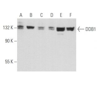
DDB1 Antibody (E-11): sc-376860
- DDB1 Antibody (E-11) is a mouse monoclonal IgG1 κ DDB1 antibody, cited in 7 publications, provided at 200 µg/ml
- specific for an epitope mapping between amino acids 9-45 near the N-terminus of DDB1 of human origin
- DDB1 Antibody (E-11) is recommended for detection of DDB1 of mouse, rat, human and origin by WB, IP, IF, IHC(P) and ELISA; also reactive with additional species, including and equine, canine, bovine, porcine and avian
- Anti-DDB1 Antibody (E-11) is available conjugated to agarose for IP; HRP for WB, IHC(P) and ELISA; and to either phycoerythrin or FITC for IF, IHC(P) and FCM
- also available conjugated to Alexa Fluor® 488, Alexa Fluor® 546, Alexa Fluor® 594 or Alexa Fluor® 647 for WB (RGB), IF, IHC(P) and FCM, and for use with RGB fluorescent imaging systems, such as iBright™ FL1000, FluorChem™, Typhoon, Azure and other comparable systems
- also available conjugated to Alexa Fluor® 680 or Alexa Fluor® 790 for WB (NIR), IF and FCM; for use with Near-Infrared (NIR) detection systems, such as LI-COR®Odyssey®, iBright™ FL1000, FluorChem™, Typhoon, Azure and other comparable systems
- m-IgG Fc BP-HRP is the preferred secondary detection reagent for DDB1 Antibody (E-11) for WB and IHC(P) applications. This reagent is now offered in a bundle with DDB1 Antibody (E-11) (see ordering information below).

QUICK LINKS
DDB1 Antibody (E-11) is a mouse monoclonal IgG1 kappa light chain antibody that detects DDB1 protein of mouse, rat, and human origin by western blotting (WB), immunoprecipitation (IP), immunofluorescence (IF), immunohistochemistry, and enzyme-linked immunosorbent assay (ELISA). anti-DDB1 antibody (E-11) is available in both non-conjugated and various conjugated forms, including agarose, horseradish peroxidase (HRP), phycoerythrin (PE), fluorescein isothiocyanate (FITC), and multiple Alexa Fluor® conjugates. DDB1, part of the damaged DNA binding protein complex, plays a crucial role in the cellular response to DNA damage, particularly damage caused by ultraviolet light. DDB1 is essential for the recognition and repair of DNA lesions, specifically cyclobutane pyrimidine dimers, which are formed when DNA is exposed to UV radiation. DDB1′s ability to bind to these damaged sites is vital for initiating the nucleotide excision repair pathway, thereby preventing mutations that could lead to skin cancer and other genetic disorders. Mutations in the DDB2 gene, which encodes the other subunit of the DDB complex, are linked to xeroderma pigmentosum group E, a condition characterized by extreme sensitivity to sunlight and a predisposition to skin malignancies. Additionally, DDB1′s interaction with the hepatitis B virus X protein suggests a potential role in viral pathogenesis, highlighting DDB1′s importance not only in DNA repair but also in viral infections.
Alexa Fluor® is a trademark of Molecular Probes Inc., OR., USA
LI-COR® and Odyssey® are registered trademarks of LI-COR Biosciences
DDB1 Antibody (E-11) References:
- Studies of the murine DDB1 and DDB2 genes. | Zolezzi, F. and Linn, S. 2000. Gene. 245: 151-9. PMID: 10713455
- Human damage-specific DNA-binding protein p48. Characterization of XPE mutations and regulation following UV irradiation. | Nichols, AF., et al. 2000. J Biol Chem. 275: 21422-8. PMID: 10777490
- Dissociation of DDB1-binding and transactivation properties of the hepatitis B virus X protein. | Wentz, MJ., et al. 2000. Virus Res. 68: 87-92. PMID: 10930665
- Identification of potential mRNA biomarkers in peripheral blood lymphocytes for human exposure to ionizing radiation. | Amundson, SA., et al. 2000. Radiat Res. 154: 342-6. PMID: 11012342
- Damaged DNA-binding protein DDB stimulates the excision of cyclobutane pyrimidine dimers in vitro in concert with XPA and replication protein A. | Wakasugi, M., et al. 2001. J Biol Chem. 276: 15434-40. PMID: 11278856
- The functions of the HIV1 protein Vpr and its action through the DCAF1.DDB1.Cullin4 ubiquitin ligase. | Casey, L., et al. 2010. Cytokine. 51: 1-9. PMID: 20347598
- HBx affects CUL4-DDB1 function in both positive and negative manners. | Guo, L., et al. 2014. Biochem Biophys Res Commun. 450: 1492-7. PMID: 25019988
- Expression of DDB1 is associated with subtypes of epithelial ovarian cancer and predicts clinical outcomes. | Shan, Y., et al. 2023. Tissue Cell. 82: 102072. PMID: 36934683
- Chromosomal localization and cDNA cloning of the genes (DDB1 and DDB2) for the p127 and p48 subunits of a human damage-specific DNA binding protein. | Dualan, R., et al. 1995. Genomics. 29: 62-9. PMID: 8530102
- Mutations specific to the xeroderma pigmentosum group E Ddb- phenotype. | Nichols, AF., et al. 1996. J Biol Chem. 271: 24317-20. PMID: 8798680
- The V protein of the paramyxovirus SV5 interacts with damage-specific DNA binding protein. | Lin, GY., et al. 1998. Virology. 249: 189-200. PMID: 9740790
- Refined mapping of the gene encoding the p127 kDa UV-damaged DNA-binding protein (DDB1) within 11q12-q13.1 and its exclusion in Best's vitelliform macular dystrophy. | Stöhr, H., et al. 1998. Eur J Hum Genet. 6: 400-5. PMID: 9781049
Ordering Information
| Product Name | Catalog # | UNIT | Price | Qty | FAVORITES | |
DDB1 Antibody (E-11) | sc-376860 | 200 µg/ml | $322.00 | |||
DDB1 Antibody (E-11): m-IgG Fc BP-HRP Bundle | sc-552123 | 200 µg Ab; 10 µg BP | $361.00 | |||
DDB1 Antibody (E-11) AC | sc-376860 AC | 500 µg/ml, 25% agarose | $424.00 | |||
DDB1 Antibody (E-11) HRP | sc-376860 HRP | 200 µg/ml | $322.00 | |||
DDB1 Antibody (E-11) FITC | sc-376860 FITC | 200 µg/ml | $336.00 | |||
DDB1 Antibody (E-11) PE | sc-376860 PE | 200 µg/ml | $349.00 | |||
DDB1 Antibody (E-11) Alexa Fluor® 488 | sc-376860 AF488 | 200 µg/ml | $364.00 | |||
DDB1 Antibody (E-11) Alexa Fluor® 546 | sc-376860 AF546 | 200 µg/ml | $364.00 | |||
DDB1 Antibody (E-11) Alexa Fluor® 594 | sc-376860 AF594 | 200 µg/ml | $364.00 | |||
DDB1 Antibody (E-11) Alexa Fluor® 647 | sc-376860 AF647 | 200 µg/ml | $364.00 | |||
DDB1 Antibody (E-11) Alexa Fluor® 680 | sc-376860 AF680 | 200 µg/ml | $364.00 | |||
DDB1 Antibody (E-11) Alexa Fluor® 790 | sc-376860 AF790 | 200 µg/ml | $364.00 | |||
DDB1 (E-11) Neutralizing Peptide | sc-376860 P | 100 µg/0.5 ml | $69.00 |