")
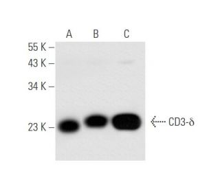
: sc-137137. Western blot analysis of CD3-δ expression in Jurkat (A), HuT 78 (B) and CCRF-CEM (C) whole cell lysates.")
 Alexa Fluor® 488: sc-137137 AF488. Direct fluorescent western blot analysis of CD3-δ expression in Jurkat (A) and CCRF-CEM (B) whole cell lysates. Blocked with UltraCruz® Blocking Reagent: sc-516214. Cruz Marker™ Molecular Weight Standards detected with Cruz Marker™ MW Tag-Alexa Fluor® 647: sc-516791.")
: sc-137137. Western blot analysis of CD3-δ expression in CCRF-CEM (A) and Jurkat (B) whole cell lysates. Detection reagent used: m-IgGκ BP-HRP: sc-516102.")
CD3-δ Antibody (F-1): sc-137137
- CD3-δ Antibody (F-1) is a mouse monoclonal IgG2a κ CD3-δ antibody, cited in 3 publications, provided at 200 µg/ml
- raised against amino acids 1-171 representing full length CD3-δ of human origin
- CD3-delta Antibody (F-1) is recommended for detection of CD3-δ of human origin by WB, IP, IF and ELISA
- Anti-CD3-delta Antibody (F-1) is available conjugated to agarose for IP; HRP for WB, IHC(P) and ELISA; and to either phycoerythrin or FITC for IF, IHC(P) and FCM
- also available conjugated to Alexa Fluor® 488, Alexa Fluor® 546, Alexa Fluor® 594 or Alexa Fluor® 647 for WB (RGB), IF, IHC(P) and FCM, and for use with RGB fluorescent imaging systems, such as iBright™ FL1000, FluorChem™, Typhoon, Azure and other comparable systems
- also available conjugated to Alexa Fluor® 680 or Alexa Fluor® 790 for WB (NIR), IF and FCM; for use with Near-Infrared (NIR) detection systems, such as LI-COR®Odyssey®, iBright™ FL1000, FluorChem™, Typhoon, Azure and other comparable systems
- m-IgG Fc BP-HRP, m-IgG2a BP-HRP and m-IgGκ BP-HRP are the preferred secondary detection reagents for CD3-δ Antibody (F-1) for WB applications. These reagents are now offered in bundles with CD3-δ Antibody (F-1) (see ordering information below).
CD3-δ Antibody (F-1) is a mouse monoclonal IgG2a kappa light chain antibody that detects CD3-delta of human origin by western blotting (WB), immunoprecipitation (IP), immunofluorescence (IF), and enzyme-linked immunosorbent assay (ELISA). Anti-CD3-delta antibody (F-1) is available in both non-conjugated and various conjugated forms, including agarose, horseradish peroxidase (HRP), phycoerythrin (PE), fluorescein isothiocyanate (FITC), and multiple Alexa Fluor® conjugates. CD3-delta plays a crucial role in the immune response as part of the T cell receptor (TCR) complex, which is essential for T cell activation and signaling. Located on the surface of T cells, CD3-delta is one of the five invariant polypeptide chains that form the CD3 complex, which is vital for the transduction of signals upon antigen recognition. This complex not only aids in the activation of T cells but also ensures that the immune system can effectively respond to pathogens. CD3-delta, along with its associated chains, allows for the formation of functional TCRs that can recognize a diverse array of antigens, making CD3-delta a key player in adaptive immunity. The intricate structure of the CD3 complex, including the immunoreceptor tyrosine-based activation motifs (ITAMs) present in the gamma, epsilon, and delta chains, facilitates the recruitment of signaling molecules, thereby amplifying the immune response. Understanding CD3-delta and its interactions within the TCR complex is essential for developing targeted therapies in immunology and cancer treatment.
Alexa Fluor® is a trademark of Molecular Probes Inc., OR., USA
LI-COR® and Odyssey® are registered trademarks of LI-COR Biosciences
CD3-δ Antibody (F-1) References:
- Role of CD3 delta in surface expression of the TCR/CD3 complex and in activation for killing analyzed with a CD3 delta-negative cytotoxic T lymphocyte variant. | Buferne, M., et al. 1992. J Immunol. 148: 657-64. PMID: 1530953
- Structure, assembly and intracellular transport of the T cell receptor for antigen. | Exley, M., et al. 1991. Semin Immunol. 3: 283-97. PMID: 1686832
- A conformational epitope expressed upon association of CD3-epsilon with either CD3-delta or CD3-gamma is the main target for recognition by anti-CD3 monoclonal antibodies. | Salmerón, A., et al. 1991. J Immunol. 147: 3047-52. PMID: 1717585
- The CD3-gamma and CD3-delta subunits of the T cell antigen receptor can be expressed within distinct functional TCR/CD3 complexes. | Alarcón, B., et al. 1991. EMBO J. 10: 903-12. PMID: 1826255
- Signal transduction by the T cell antigen receptor. | Weiss, A., et al. 1991. Semin Immunol. 3: 313-24. PMID: 1839225
- Human T cell generation is restored in CD3δ severe combined immunodeficiency through adenine base editing. | McAuley, GE., et al. 2023. Cell. 186: 1398-1416.e23. PMID: 36944331
- Signal transduction. Zapping tandem SH2 domains. | Weiss, A. 1995. Nature. 377: 17-8. PMID: 7659151
- Molecular basis for interaction of the protein tyrosine kinase ZAP-70 with the T-cell receptor. | Hatada, MH., et al. 1995. Nature. 377: 32-8. PMID: 7659156
- Binding of ZAP-70 to phosphorylated T-cell receptor zeta and eta enhances its autophosphorylation and generates specific binding sites for SH2 domain-containing proteins. | Neumeister, EN., et al. 1995. Mol Cell Biol. 15: 3171-8. PMID: 7760813
- Different cytoplasmic structure of the CD3 zeta family dimer modulates the activation signal and function of T cells. | Aoe, T., et al. 1994. Int Immunol. 6: 1671-9. PMID: 7865460
- The role of protein tyrosine kinases and protein tyrosine phosphatases in T cell antigen receptor signal transduction. | Chan, AC., et al. 1994. Annu Rev Immunol. 12: 555-92. PMID: 8011291
- Targeted disruption of the CD3 eta locus causes high lethality in mice: modulation of Oct-1 transcription on the opposite strand. | Ohno, H., et al. 1994. EMBO J. 13: 1157-65. PMID: 8131747
Ordering Information
| Product Name | Catalog # | UNIT | Price | Qty | FAVORITES | |
CD3-δ Antibody (F-1) | sc-137137 | 200 µg/ml | $316.00 | |||
CD3-δ Antibody (F-1): m-IgG Fc BP-HRP Bundle | sc-528867 | 200 µg Ab; 10 µg BP | $354.00 | |||
CD3-δ Antibody (F-1): m-IgGκ BP-HRP Bundle | sc-521435 | 200 µg Ab, 40 µg BP | $354.00 | |||
CD3-δ Antibody (F-1): m-IgG2a BP-HRP Bundle | sc-547195 | 200 µg Ab; 10 µg BP | $354.00 | |||
CD3-δ Antibody (F-1) AC | sc-137137 AC | 500 µg/ml, 25% agarose | $416.00 | |||
CD3-δ Antibody (F-1) HRP | sc-137137 HRP | 200 µg/ml | $316.00 | |||
CD3-δ Antibody (F-1) FITC | sc-137137 FITC | 200 µg/ml | $330.00 | |||
CD3-δ Antibody (F-1) PE | sc-137137 PE | 200 µg/ml | $343.00 | |||
CD3-δ Antibody (F-1) Alexa Fluor® 488 | sc-137137 AF488 | 200 µg/ml | $357.00 | |||
CD3-δ Antibody (F-1) Alexa Fluor® 546 | sc-137137 AF546 | 200 µg/ml | $357.00 | |||
CD3-δ Antibody (F-1) Alexa Fluor® 594 | sc-137137 AF594 | 200 µg/ml | $357.00 | |||
CD3-δ Antibody (F-1) Alexa Fluor® 647 | sc-137137 AF647 | 200 µg/ml | $357.00 | |||
CD3-δ Antibody (F-1) Alexa Fluor® 680 | sc-137137 AF680 | 200 µg/ml | $357.00 | |||
CD3-δ Antibody (F-1) Alexa Fluor® 790 | sc-137137 AF790 | 200 µg/ml | $357.00 |