")
ATR Anticuerpo (C-1): sc-515173
- ATR Anticuerpo C-1 es un monoclonal de ratón IgG1 κ ATR Anticuerpo, ver las 50 publicaciones, proporcionado como 200 µg/ml
- específico para un epítopo localizado entre los amino ácidos 2596-2621 cerca del C-terminus de ATR oe orgiendf humanorigin
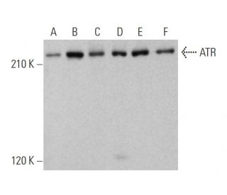
- ATR Anticuerpo (C-1) es recomendado para detectar ATR de mouse, rat y human origen, mediante WB, IP, IF y ELISA
- ATR Anticuerpo (C-1) es disponible conjugado a agarosa para IP; HRP para WB, IHC(P) y ELISA; y tanto a phycoerythrin como a FITC para IF, IHC(P) y FCM
- también disponible conjugado a Alexa Fluor® 488, Alexa Fluor® 546, Alexa Fluor® 594 o Alexa Fluor® 647 para WB (RGB), IF, IHC (P) y FCM
- también disponible conjugado a Alexa Fluor® 680 o Alexa Fluor® 790 para WB (NIR), IF y FCM
- m-IgG Fc BP-HRP, 1 BP-HRP">m-IgG1 BP-HRP y m-IgGκ BP-HRP son los reactivos de detección secundarios preferidos para ATR Anticuerpo (C-1) para aplicaciones WB. Estos reactivos se ofrecen ahora en paquetes con ATR Anticuerpo (C-1)(véase la información de pedido más abajo).

ENLACES RÁPIDOS
VER TAMBIÉN ....
El anticuerpo ATR (C-1) es un anticuerpo monoclonal IgG1 de ratón de cadena ligera kappa que detecta la proteína ATR de origen de ratón, rata y humano mediante western blotting (WB), inmunoprecipitación (IP), inmunofluorescencia (IF) y ensayo inmunoenzimático (ELISA). El anticuerpo anti-ATR (C-1) está disponible tanto en forma no conjugada como en varias formas conjugadas, incluyendo agarosa, peroxidasa de rábano picante (HRP), ficoeritrina (PE), isotiocianato de fluoresceína (FITC) y múltiples conjugados Alexa Fluor®. ATR, también conocida como ataxia-telangiectasia y proteína relacionada con Rad3, desempeña un papel crucial en la respuesta celular al daño del ADN, en particular en la activación de los puntos de control del ciclo celular. ATR fosforila proteínas clave implicadas en la respuesta al daño del ADN, como la quinasa de punto de control CHK1 y el supresor tumoral BRCA1, lo que hace que ATR sea vital para mantener la estabilidad genómica. ATR se localiza principalmente en el núcleo, donde forma focos intranucleares en respuesta al daño del ADN o al estrés de replicación, lo que pone de relieve la importancia de ATR en los mecanismos de reparación celular. ATR es esencial para el desarrollo embrionario temprano, lo que subraya la importancia de ATR tanto en la función celular normal como en la prevención de enfermedades asociadas a la inestabilidad genómica.
Alexa Fluor® es una marca registrada de Molecular Probes Inc., OR., USA
REIVEW LI-COR® y Odyssey® son marcas registradas de LI-COR Biosciences.
ATR Anticuerpo (C-1) Referencias:
- Asociación molecular entre ATR y dos componentes del complejo de remodelación y desacetilación de nucleosomas, HDAC2 y CHD4. | Schmidt, DR. and Schreiber, SL. 1999. Biochemistry. 38: 14711-7. PMID: 10545197
- La ATR es una proteína cinasa activada por el ADN, sensible a la cafeína, con una especificidad de sustrato distinta de la DNA-PK. | Hall-Jackson, CA., et al. 1999. Oncogene. 18: 6707-13. PMID: 10597277
- Una vía ATR/CHK1 sobreactivada es responsable de la acumulación G2 prolongada en células AT irradiadas. | Wang, X., et al. 2003. J Biol Chem. 278: 30869-74. PMID: 12791699
- MSH2 y ATR forman un módulo de señalización y regulan dos ramas de la respuesta al daño por metilación del ADN. | Wang, Y. and Qin, J. 2003. Proc Natl Acad Sci U S A. 100: 15387-92. PMID: 14657349
- Una nueva actividad proteica media la unión al ADN de un complejo ATR-ATRIP. | Bomgarden, RD., et al. 2004. J Biol Chem. 279: 13346-53. PMID: 14724280
- La vía ATR-p53 se suprime en linfocitos normales y malignos no ciclantes. | Jones, GG., et al. 2004. Oncogene. 23: 1911-21. PMID: 14755251
- La inhibición de ATR conduce a una mayor sensibilidad a la hipoxia/reoxigenación. | Hammond, EM., et al. 2004. Cancer Res. 64: 6556-62. PMID: 15374968
- ATR ataca la envoltura nuclear. | Smolka, MB. and Lammerding, J. 2023. Mol Cell. 83: 3588-3590. PMID: 37863026
- La pérdida de FBXW7 sensibiliza a las células a la inhibición de ATR a través de la catástrofe mitótica inducida. | O'Brien, S., et al. 2023. Cancer Res Commun. 3: 2596-2607. PMID: 38032106
- Primer estudio en humanos del inhibidor de la ataxia telangiectasia y relacionado con Rad3 (ATR) Tuvusertib (M1774) como monoterapia en pacientes con tumores sólidos. | Yap, TA., et al. 2024. Clin Cancer Res. 30: 2057-2067. PMID: 38407317
- Clonación de ADNc y cartografía génica de una proteína candidata del punto de control del ciclo celular humano. | Cimprich, KA., et al. 1996. Proc Natl Acad Sci U S A. 93: 2850-5. PMID: 8610130
- Las proteínas quinasas Atr y Atm se asocian a sitios diferentes a lo largo de los cromosomas emparejados meióticamente. | Keegan, KS., et al. 1996. Genes Dev. 10: 2423-37. PMID: 8843195
Información sobre pedidos
| Nombre del producto | Número de catálogo | UNIDAD | Precio | CANTIDAD | Favoritos | |
ATR Anticuerpo (C-1) | sc-515173 | 200 µg/ml | $322.00 | |||
Paquete de ATR (C-1): m-IgG Fc BP-HRP | sc-531302 | 200 µg Ab; 10 µg BP | $361.00 | |||
Paquete de ATR (C-1): m-IgGκ BP-HRP | sc-524960 | 200 µg Ab, 40 µg BP | $361.00 | |||
Paquete de ATR (C-1): m-IgG1 BP-HRP | sc-544473 | 200 µg Ab; 20 µg BP | $361.00 | |||
ATR Anticuerpo (C-1) AC | sc-515173 AC | 500 µg/ml, 25% agarose | $424.00 | |||
ATR Anticuerpo (C-1) HRP | sc-515173 HRP | 200 µg/ml | $322.00 | |||
ATR Anticuerpo (C-1) FITC | sc-515173 FITC | 200 µg/ml | $336.00 | |||
ATR Anticuerpo (C-1) PE | sc-515173 PE | 200 µg/ml | $349.00 | |||
ATR Anticuerpo (C-1) Alexa Fluor® 488 | sc-515173 AF488 | 200 µg/ml | $364.00 | |||
ATR Anticuerpo (C-1) Alexa Fluor® 546 | sc-515173 AF546 | 200 µg/ml | $364.00 | |||
ATR Anticuerpo (C-1) Alexa Fluor® 594 | sc-515173 AF594 | 200 µg/ml | $364.00 | |||
ATR Anticuerpo (C-1) Alexa Fluor® 647 | sc-515173 AF647 | 200 µg/ml | $364.00 | |||
ATR Anticuerpo (C-1) Alexa Fluor® 680 | sc-515173 AF680 | 200 µg/ml | $364.00 | |||
ATR Anticuerpo (C-1) Alexa Fluor® 790 | sc-515173 AF790 | 200 µg/ml | $364.00 | |||
ATR (C-1) péptido neutralizante | sc-515173 P | 100 µg/0.5 ml | $69.00 |