")
XPA Antibody (B-1): sc-28353
- XPA Antibody (B-1) is a mouse monoclonal IgG1 κ XPA antibody, cited in 26 publications, provided at 200 µg/ml
- raised against amino acids 1-273 representing full length XPA of human origin
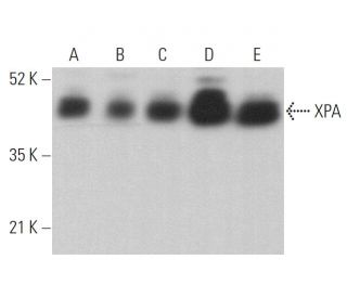
- XPA Antibody (B-1) is recommended for detection of XPA of mouse, rat and human origin by WB, IP, IF, IHC(P) and ELISA
- Anti-XPA Antibody (B-1) is available conjugated to agarose for IP; HRP for WB, IHC(P) and ELISA; and to either phycoerythrin or FITC for IF, IHC(P) and FCM
- also available conjugated to Alexa Fluor® 488, Alexa Fluor® 546, Alexa Fluor® 594 or Alexa Fluor® 647 for WB (RGB), IF, IHC(P) and FCM, and for use with RGB fluorescent imaging systems, such as iBright™ FL1000, FluorChem™, Typhoon, Azure and other comparable systems
- also available conjugated to Alexa Fluor® 680 or Alexa Fluor® 790 for WB (NIR), IF and FCM; for use with Near-Infrared (NIR) detection systems, such as LI-COR®Odyssey®, iBright™ FL1000, FluorChem™, Typhoon, Azure and other comparable systems
- m-IgG Fc BP-HRP, m-IgG1 BP-HRP and m-IgGκ BP-HRP are the preferred secondary detection reagents for XPA Antibody (B-1) for WB and IHC(P) applications. These reagents are now offered in bundles with XPA Antibody (B-1) (see ordering information below).

QUICK LINKS
SEE ALSO...
XPA Antibody (B-1) is a mouse monoclonal IgG1 kappa light chain antibody that detects XPA protein of mouse, rat, and human origin by western blotting (WB), immunoprecipitation (IP), immunofluorescence (IF), immunohistochemistry, and enzyme-linked immunosorbent assay (ELISA). anti-XPA antibody (B-1) is available in both non-conjugated and various conjugated forms, including agarose, horseradish peroxidase (HRP), phycoerythrin (PE), fluorescein isothiocyanate (FITC), and multiple Alexa Fluor® conjugates. XPA protein plays a critical role in the nucleotide excision repair (NER) pathway, which is essential for repairing DNA damage caused by ultraviolet (UV) light. This function is particularly important as deficiencies in NER can lead to xeroderma pigmentosum, a genetic disorder that significantly increases the risk of skin cancer due to the accumulation of unrepaired DNA lesions. XPA is responsible for coordinating the assembly of the preincision complex necessary for recognizing and excising damaged DNA, and XPA exhibits a unique ability to bind to both single-stranded and double-stranded DNA, enhancing efficiency in damage recognition. Furthermore, XPA′s ability to form homodimers in the absence of DNA and transition to a more effective dimeric form upon binding to DNA underscores the regulatory mechanisms that control activity in response to DNA damage. XPA′s importance in maintaining genomic integrity highlights potential as a target for therapeutic interventions in conditions associated with impaired DNA repair mechanisms.
Alexa Fluor® is a trademark of Molecular Probes Inc., OR., USA
LI-COR® and Odyssey® are registered trademarks of LI-COR Biosciences
XPA Antibody (B-1) References:
- The relative expression of mutated XPB genes results in xeroderma pigmentosum/Cockayne's syndrome or trichothiodystrophy cellular phenotypes. | Riou, L., et al. 1999. Hum Mol Genet. 8: 1125-33. PMID: 10332046
- Photobiologic and photoimmunologic characteristics of XPA gene-deficient mice. | Horio, T., et al. 2001. J Investig Dermatol Symp Proc. 6: 58-63. PMID: 11764287
- Xeroderma pigmentosum complementation group A protein (XPA) modulates RPA-DNA interactions via enhanced complex stability and inhibition of strand separation activity. | Patrick, SM. and Turchi, JJ. 2002. J Biol Chem. 277: 16096-101. PMID: 11859086
- Cooperative interaction of human XPA stabilizes and enhances specific binding of XPA to DNA damage. | Liu, Y., et al. 2005. Biochemistry. 44: 7361-8. PMID: 15882075
- Opposing effects of the UV lesion repair protein XPA and UV bypass polymerase eta on ATR checkpoint signaling. | Bomgarden, RD., et al. 2006. EMBO J. 25: 2605-14. PMID: 16675950
- ATR-dependent checkpoint modulates XPA nuclear import in response to UV irradiation. | Wu, X., et al. 2007. Oncogene. 26: 757-64. PMID: 16862173
- XPA deficiency affects the ubiquitin-proteasome system function. | de Sousa Leal, AM., et al. 2020. DNA Repair (Amst). 94: 102937. PMID: 32693352
- Lesion recognition by XPC, TFIIH and XPA in DNA excision repair. | Kim, J., et al. 2023. Nature. 617: 170-175. PMID: 37076618
- Cross-species investigation into the requirement of XPA for nucleotide excision repair. | Kose, C., et al. 2024. Nucleic Acids Res. 52: 677-689. PMID: 37994737
- High incidence of ultraviolet-B-or chemical-carcinogen-induced skin tumours in mice lacking the xeroderma pigmentosum group A gene. | Nakane, H., et al. 1995. Nature. 377: 165-8. PMID: 7675085
- Identification of a damaged-DNA binding domain of the XPA protein. | Kuraoka, I., et al. 1996. Mutat Res. 362: 87-95. PMID: 8538652
- Separation of protein factors that correct the defects in the seven complementation groups of xeroderma pigmentosum cells. | Tateishi, S., et al. 1995. J Biochem. 118: 819-24. PMID: 8576098
Ordering Information
| Product Name | Catalog # | UNIT | Price | Qty | FAVORITES | |
XPA Antibody (B-1) | sc-28353 | 200 µg/ml | $322.00 | |||
XPA Antibody (B-1): m-IgG Fc BP-HRP Bundle | sc-528432 | 200 µg Ab; 10 µg BP | $361.00 | |||
XPA Antibody (B-1): m-IgGκ BP-HRP Bundle | sc-520807 | 200 µg Ab, 40 µg BP | $361.00 | |||
XPA Antibody (B-1): m-IgG1 BP-HRP Bundle | sc-542931 | 200 µg Ab; 20 µg BP | $361.00 | |||
XPA Antibody (B-1) AC | sc-28353 AC | 500 µg/ml, 25% agarose | $424.00 | |||
XPA Antibody (B-1) HRP | sc-28353 HRP | 200 µg/ml | $322.00 | |||
XPA Antibody (B-1) FITC | sc-28353 FITC | 200 µg/ml | $336.00 | |||
XPA Antibody (B-1) PE | sc-28353 PE | 200 µg/ml | $349.00 | |||
XPA Antibody (B-1) Alexa Fluor® 488 | sc-28353 AF488 | 200 µg/ml | $364.00 | |||
XPA Antibody (B-1) Alexa Fluor® 546 | sc-28353 AF546 | 200 µg/ml | $364.00 | |||
XPA Antibody (B-1) Alexa Fluor® 594 | sc-28353 AF594 | 200 µg/ml | $364.00 | |||
XPA Antibody (B-1) Alexa Fluor® 647 | sc-28353 AF647 | 200 µg/ml | $364.00 | |||
XPA Antibody (B-1) Alexa Fluor® 680 | sc-28353 AF680 | 200 µg/ml | $364.00 | |||
XPA Antibody (B-1) Alexa Fluor® 790 | sc-28353 AF790 | 200 µg/ml | $364.00 |