")
Visual Arrestin Antibody (E-3): sc-166383
- Visual Arrestin Antibody (E-3) is a mouse monoclonal IgG2b κ Visual Arrestin antibody, cited in 8 publications, provided at 200 µg/ml
- raised against amino acids 316-405 mapping at the C-terminus of Visual Arrestin of human origin
- Visual Arrestin Antibody (E-3) is recommended for detection of Visual Arrestin of mouse, rat and human origin by WB, IP, IF, IHC(P) and ELISA
- Anti-Visual Arrestin Antibody (E-3) is available conjugated to agarose for IP; HRP for WB, IHC(P) and ELISA; and to either phycoerythrin or FITC for IF, IHC(P) and FCM
- also available conjugated to Alexa Fluor® 488, Alexa Fluor® 546, Alexa Fluor® 594 or Alexa Fluor® 647 for WB (RGB), IF, IHC(P) and FCM, and for use with RGB fluorescent imaging systems, such as iBright™ FL1000, FluorChem™, Typhoon, Azure and other comparable systems
- also available conjugated to Alexa Fluor® 680 or Alexa Fluor® 790 for WB (NIR), IF and FCM; for use with Near-Infrared (NIR) detection systems, such as LI-COR®Odyssey®, iBright™ FL1000, FluorChem™, Typhoon, Azure and other comparable systems
- m-IgG2b BP-HRP and m-IgGκ BP-HRP are the preferred secondary detection reagents for Visual Arrestin Antibody (E-3) for WB and IHC(P) applications. These reagents are now offered in bundles with Visual Arrestin Antibody (E-3) (see ordering information below).

QUICK LINKS
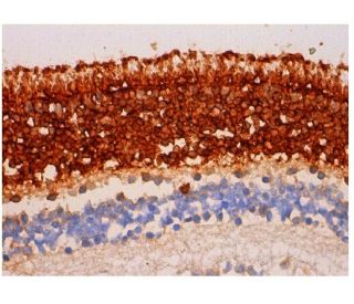
Visual Arrestin Antibody (E-3) is a mouse monoclonal IgG2b kappa light chain antibody designed to detect Visual Arrestin, also known as Arrestin 1, retinal S-antigen, or S-Arrestin, in human, mouse, and rat species through applications such as western blotting (WB), immunoprecipitation (IP), immunofluorescence (IF), immunohistochemistry with paraffin-embedded sections (IHCP), and enzyme-linked immunosorbent assay (ELISA). Visual Arrestin monoclonal antibody (E-3) is raised against amino acids 316-405 at the C-terminus of the human Visual Arrestin protein, ensuring effective binding for reliable detection. Visual Arrestin plays a vital role in the phototransduction pathway by binding to phosphorylated rhodopsin, thereby terminating the light signal and preventing continuous activation of G protein-coupled receptors. Structurally, Visual Arrestin consists of an N-terminal domain and a C-terminal domain that form a compact configuration essential for interaction with rhodopsin and other signaling proteins. This structural arrangement enables Visual Arrestin to effectively regulate receptor activity, ensuring precise control of visual signal transduction and maintaining accurate visual perception. Additionally, Visual Arrestin is localized in the outer segments of retinal photoreceptor cells and the pineal gland, where Visual Arrestin contributes to the regulation of circadian rhythms, highlighting importance in both vision and overall physiological processes. The specific cellular localization of Visual Arrestin is crucial for function in deactivating rhodopsin, thereby maintaining cellular homeostasis and proper visual function. Furthermore, Visual Arrestin serves as a major pathogenic autoantigen in inflammatory eye diseases such as uveoretinitis and Oguchi disease, a rare form of night blindness, underscoring significance in ocular health and disease. anti-Visual Arrestin antibody (E-3) facilitates detailed studies of structure and function, supporting research into visual signal transduction, retinal disorders, and autoimmune conditions.
Alexa Fluor® is a trademark of Molecular Probes Inc., OR., USA
LI-COR® and Odyssey® are registered trademarks of LI-COR Biosciences
Visual Arrestin Antibody (E-3) References:
- Gene analysis and evaluation of the single founder effect in Japanese patients with Oguchi disease. | Saga, M., et al. 2004. Jpn J Ophthalmol. 48: 350-2. PMID: 15295660
- Induction of experimental autoimmune uveoretinitis in Lewis rats with purified recombinant human retinal S-antigen fusion protein. | Roberts, AJ., et al. 1992. Eur J Immunol. 22: 951-6. PMID: 1551407
- Structural organization of the human S-antigen gene. cDNA, amino acid, intron, exon, promoter, in vitro transcription, retina, and pineal gland. | Yamaki, K., et al. 1990. J Biol Chem. 265: 20757-62. PMID: 2249983
- Analysis of antigenic determinants of retinal S-antigen with monoclonal antibodies. | Banga, JP., et al. 1988. Invest Ophthalmol Vis Sci. 29: 12-21. PMID: 2447030
- Regulation of rhodopsin dephosphorylation by arrestin. | Palczewski, K., et al. 1989. J Biol Chem. 264: 15770-3. PMID: 2550422
Ordering Information
| Product Name | Catalog # | UNIT | Price | Qty | FAVORITES | |
Visual Arrestin Antibody (E-3) | sc-166383 | 200 µg/ml | $322.00 | |||
Visual Arrestin Antibody (E-3): m-IgGκ BP-HRP Bundle | sc-521583 | 200 µg Ab, 40 µg BP | $361.00 | |||
Visual Arrestin Antibody (E-3): m-IgG2b BP-HRP Bundle | sc-548932 | 200 µg Ab; 10 µg BP | $361.00 | |||
Visual Arrestin Antibody (E-3) AC | sc-166383 AC | 500 µg/ml, 25% agarose | $424.00 | |||
Visual Arrestin Antibody (E-3) HRP | sc-166383 HRP | 200 µg/ml | $322.00 | |||
Visual Arrestin Antibody (E-3) FITC | sc-166383 FITC | 200 µg/ml | $336.00 | |||
Visual Arrestin Antibody (E-3) PE | sc-166383 PE | 200 µg/ml | $349.00 | |||
Visual Arrestin Antibody (E-3) Alexa Fluor® 488 | sc-166383 AF488 | 200 µg/ml | $364.00 | |||
Visual Arrestin Antibody (E-3) Alexa Fluor® 546 | sc-166383 AF546 | 200 µg/ml | $364.00 | |||
Visual Arrestin Antibody (E-3) Alexa Fluor® 594 | sc-166383 AF594 | 200 µg/ml | $364.00 | |||
Visual Arrestin Antibody (E-3) Alexa Fluor® 647 | sc-166383 AF647 | 200 µg/ml | $364.00 | |||
Visual Arrestin Antibody (E-3) Alexa Fluor® 680 | sc-166383 AF680 | 200 µg/ml | $364.00 | |||
Visual Arrestin Antibody (E-3) Alexa Fluor® 790 | sc-166383 AF790 | 200 µg/ml | $364.00 |