")
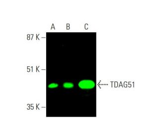
TDAG51 Antibody (RN-6E2): sc-23866
- TDAG51 Antibody (RN-6E2) is a mouse monoclonal IgG2a κ TDAG51 antibody, cited in 38 publications, provided at 200 µg/ml
- raised against TDAG51 of human origin
- TDAG51 Antibody (RN-6E2) is recommended for detection of TDAG51 of mouse, rat and human origin by WB, IP, IF, IHC(P) and FCM
- Anti-TDAG51 Antibody (RN-6E2) is available conjugated to agarose for IP; HRP for WB, IHC(P) and ELISA; and to either phycoerythrin or FITC for IF, IHC(P) and FCM
- also available conjugated to Alexa Fluor® 488, Alexa Fluor® 546, Alexa Fluor® 594 or Alexa Fluor® 647 for WB (RGB), IF, IHC(P) and FCM, and for use with RGB fluorescent imaging systems, such as iBright™ FL1000, FluorChem™, Typhoon, Azure and other comparable systems
- also available conjugated to Alexa Fluor® 680 or Alexa Fluor® 790 for WB (NIR), IF and FCM; for use with Near-Infrared (NIR) detection systems, such as LI-COR®Odyssey®, iBright™ FL1000, FluorChem™, Typhoon, Azure and other comparable systems
- m-IgG Fc BP-HRP, m-IgG2a BP-HRP and m-IgGκ BP-HRP are the preferred secondary detection reagents for TDAG51 Antibody (RN-6E2) for WB and IHC(P) applications. These reagents are now offered in bundles with TDAG51 Antibody (RN-6E2) (see ordering information below).

QUICK LINKS
SEE ALSO...
TDAG51 Antibody (RN-6E2) is a mouse monoclonal IgG2a kappa light chain antibody that detects TDAG51 protein of mouse, rat, and human origin by western blotting (WB), immunoprecipitation (IP), immunofluorescence (IF), immunohistochemistry, and flow cytometry (FCM). Anti-TDAG51 antibody (RN-6E2) is available in both non-conjugated and various conjugated forms, including agarose, horseradish peroxidase (HRP), phycoerythrin (PE), fluorescein isothiocyanate (FITC), and multiple Alexa Fluor® conjugates. TDAG51, also known as Pleckstrin Homology Like Domain Family A Member 1, plays a pivotal role in regulating T cell apoptosis, which is crucial for maintaining immune homeostasis and preventing autoimmunity. Located primarily in cytoplasm and nucleus of T cells, TDAG51 is involved in signaling pathways that link T cell receptor stimulation to FAS receptor expression, a key player in apoptotic process. TDAG51 serves as a critical mediator in immune response by restoring activation-induced apoptosis in T cells that have lost FAS expression ability. TDAG51′s role in modulating apoptosis highlights its importance in immuno-surveillance and overall regulation of immune responses, making TDAG51 a valuable target for research in immunology and cancer therapy.
Alexa Fluor® is a trademark of Molecular Probes Inc., OR., USA
LI-COR® and Odyssey® are registered trademarks of LI-COR Biosciences
TDAG51 Antibody (RN-6E2) References:
- TDAG51 is not essential for Fas/CD95 regulation and apoptosis in vivo. | Rho, J., et al. 2001. Mol Cell Biol. 21: 8365-70. PMID: 11713273
- Role of perforin in lymphocyte-mediated cytolysis. | Yagita, H., et al. 1992. Adv Immunol. 51: 215-42. PMID: 1502975
- A central role of perforin in cytolysis? | Podack, ER., et al. 1991. Annu Rev Immunol. 9: 129-57. PMID: 1910674
- Perforin-dependent and -independent pathways of cytotoxicity mediated by lymphocytes. | Young, JD., et al. 1988. Immunol Rev. 103: 161-202. PMID: 3292393
- TDAG51-Deficiency Podocytes are Protected from High-Glucose-Induced Damage Through Nrf2 Activation via the AKT-GSK-3β Pathway. | Liu, C., et al. 2022. Inflammation. 45: 1520-1533. PMID: 35175494
- TDAG51 Attenuates Impaired Lipid Metabolism and Insulin Resistance in Gestational Diabetes Mellitus Through SREBP-1/ANGPTL8 Pathway. | Wu, X. and Xiao, B. 2023. Balkan Med J. 40: 175-181. PMID: 36960944
- TDAG51 promotes transcription factor FoxO1 activity during LPS-induced inflammatory responses. | Park, ES., et al. 2023. EMBO J. 42: e111867. PMID: 37203866
- Restoration of the ER stress response protein TDAG51 in hepatocytes mitigates NAFLD in mice. | Yousof, TR., et al. 2024. J Biol Chem. 300: 105655. PMID: 38237682
- Mechanism of lymphocyte-mediated cytotoxicity. | Henkart, PA. 1985. Annu Rev Immunol. 3: 31-58. PMID: 3904772
- Fas and its ligand in a general mechanism of T-cell-mediated cytotoxicity. | Hanabuchi, S., et al. 1994. Proc Natl Acad Sci U S A. 91: 4930-4. PMID: 7515183
- The Fas protein is expressed at high levels on CD4+CD8+ thymocytes and activated mature lymphocytes in normal mice but not in the lupus-prone strain, MRL lpr/lpr. | Drappa, J., et al. 1993. Proc Natl Acad Sci U S A. 90: 10340-4. PMID: 7694292
- A novel gene product that couples TCR signaling to Fas(CD95) expression in activation-induced cell death. | Park, CG., et al. 1996. Immunity. 4: 583-91. PMID: 8673705
Ordering Information
| Product Name | Catalog # | UNIT | Price | Qty | FAVORITES | |
TDAG51 Antibody (RN-6E2) | sc-23866 | 200 µg/ml | $322.00 | |||
TDAG51 Antibody (RN-6E2): m-IgG Fc BP-HRP Bundle | sc-528373 | 200 µg Ab; 10 µg BP | $361.00 | |||
TDAG51 Antibody (RN-6E2): m-IgGκ BP-HRP Bundle | sc-520735 | 200 µg Ab, 40 µg BP | $361.00 | |||
TDAG51 Antibody (RN-6E2): m-IgG2a BP-HRP Bundle | sc-547079 | 200 µg Ab; 10 µg BP | $361.00 | |||
TDAG51 Antibody (RN-6E2) AC | sc-23866 AC | 500 µg/ml, 25% agarose | $424.00 | |||
TDAG51 Antibody (RN-6E2) HRP | sc-23866 HRP | 200 µg/ml | $322.00 | |||
TDAG51 Antibody (RN-6E2) FITC | sc-23866 FITC | 200 µg/ml | $336.00 | |||
TDAG51 Antibody (RN-6E2) PE | sc-23866 PE | 200 µg/ml | $349.00 | |||
TDAG51 Antibody (RN-6E2) Alexa Fluor® 488 | sc-23866 AF488 | 200 µg/ml | $364.00 | |||
TDAG51 Antibody (RN-6E2) Alexa Fluor® 546 | sc-23866 AF546 | 200 µg/ml | $364.00 | |||
TDAG51 Antibody (RN-6E2) Alexa Fluor® 594 | sc-23866 AF594 | 200 µg/ml | $364.00 | |||
TDAG51 Antibody (RN-6E2) Alexa Fluor® 647 | sc-23866 AF647 | 200 µg/ml | $364.00 | |||
TDAG51 Antibody (RN-6E2) Alexa Fluor® 680 | sc-23866 AF680 | 200 µg/ml | $364.00 | |||
TDAG51 Antibody (RN-6E2) Alexa Fluor® 790 | sc-23866 AF790 | 200 µg/ml | $364.00 |