")
Tak1 Antibody (C-9): sc-7967
- Tak1 Antibody (C-9) is a mouse monoclonal IgG2a κ Tak1 antibody, cited in 66 publications, provided at 200 µg/ml
- raised against amino acids 1-579 representing full length Tak1 (TGFβ-activated kinase) of mouse origin
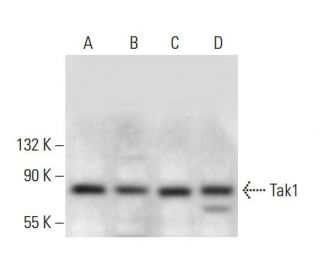
- Tak1 Antibody (C-9) is recommended for detection of Tak1 of mouse, rat and human origin by WB, IP, IF and ELISA
- Anti-Tak1 Antibody (C-9) is available conjugated to agarose for IP; HRP for WB, IHC(P) and ELISA; and to either phycoerythrin or FITC for IF, IHC(P) and FCM
- also available conjugated to Alexa Fluor® 488, Alexa Fluor® 546, Alexa Fluor® 594 or Alexa Fluor® 647 for WB (RGB), IF, IHC(P) and FCM, and for use with RGB fluorescent imaging systems, such as iBright™ FL1000, FluorChem™, Typhoon, Azure and other comparable systems
- also available conjugated to Alexa Fluor® 680 or Alexa Fluor® 790 for WB (NIR), IF and FCM; for use with Near-Infrared (NIR) detection systems, such as LI-COR®Odyssey®, iBright™ FL1000, FluorChem™, Typhoon, Azure and other comparable systems
- m-IgG Fc BP-HRP and m-IgGκ BP-HRP are the preferred secondary detection reagents for Tak1 Antibody (C-9) for WB applications. These reagents are now offered in bundles with Tak1 Antibody (C-9) (see ordering information below).

QUICK LINKS
Tak1 Antibody (C-9) is a mouse monoclonal IgG2a kappa kappa light chain antibody raised against amino acids 1-579 of mouse Tak1 (TGFβ-activated kinase 1), also known as MAP3K7 (Mitogen-Activated Protein Kinase Kinase Kinase 7). Tak1 monoclonal antibody (C-9) detects Tak1 protein of mouse, rat, and human origin by western blotting (WB), immunoprecipitation (IP), immunofluorescence (IF), and enzyme-linked immunosorbent assay (ELISA). Tak1 is a serine/threonine kinase that plays a pivotal role in mediating cellular responses to cytokines like TGFβ and interleukin-1 (IL-1), activating downstream NF-κB and MAP kinase signaling pathways essential for inflammation, immune responses, cell survival, and apoptosis. Dysregulation of Tak1 function is associated with various pathological conditions, including cancer and inflammatory diseases, making Tak1 a critical target for biomedical research. Tak1 monoclonal antibody (C-9) enables the study of key signaling pathways and their roles in disease mechanisms. Tak1 monoclonal antibody (C-9) is available in non-conjugated form and multiple conjugated forms, including agarose, HRP, PE, FITC, and several Alexa Fluor® conjugates, facilitating versatile applications in research.
Alexa Fluor® is a trademark of Molecular Probes Inc., OR., USA
LI-COR® and Odyssey® are registered trademarks of LI-COR Biosciences
Tak1 Antibody (C-9) References:
- Protein kinase cascades in meiotic and mitotic cell cycle control. | Pelech, SL., et al. 1990. Biochem Cell Biol. 68: 1297-330. PMID: 2085430
- Signal transduction from membrane to cytoplasm: growth factors and membrane-bound oncogene products increase Raf-1 phosphorylation and associated protein kinase activity. | Morrison, DK., et al. 1988. Proc Natl Acad Sci U S A. 85: 8855-9. PMID: 3057494
- Insulin-stimulated microtubule-associated protein kinase is phosphorylated on tyrosine and threonine in vivo. | Ray, LB. and Sturgill, TW. 1988. Proc Natl Acad Sci U S A. 85: 3753-7. PMID: 3287375
- TAK1 mediates neuronal pyroptosis in early brain injury after subarachnoid hemorrhage. | Xu, P., et al. 2021. J Neuroinflammation. 18: 188. PMID: 34461942
- Characterization of murine A-raf, a new oncogene related to the v-raf oncogene. | Huleihel, M., et al. 1986. Mol Cell Biol. 6: 2655-62. PMID: 3491291
- The Role of TAK1 in RANKL-Induced Osteoclastogenesis. | Jianwei, W., et al. 2022. Calcif Tissue Int. 111: 1-12. PMID: 35286417
- TAK1 deficiency promotes liver injury and tumorigenesis via ferroptosis and macrophage cGAS-STING signalling. | Su, W., et al. 2023. JHEP Rep. 5: 100695. PMID: 36968217
- TAK1 is an essential kinase for STING trafficking. | Ma, M., et al. 2023. Mol Cell. 83: 3885-3903.e5. PMID: 37832545
- KSR: a novel player in the RAS pathway. | Downward, J. 1995. Cell. 83: 831-4. PMID: 8521506
- KSR, a novel protein kinase required for RAS signal transduction. | Therrien, M., et al. 1995. Cell. 83: 879-88. PMID: 8521512
- The C. elegans ksr-1 gene encodes a novel Raf-related kinase involved in Ras-mediated signal transduction. | Sundaram, M. and Han, M. 1995. Cell. 83: 889-901. PMID: 8521513
- Identification of a member of the MAPKKK family as a potential mediator of TGF-beta signal transduction. | Yamaguchi, K., et al. 1995. Science. 270: 2008-11. PMID: 8533096
Ordering Information
| Product Name | Catalog # | UNIT | Price | Qty | FAVORITES | |
Tak1 Antibody (C-9) | sc-7967 | 200 µg/ml | $322.00 | |||
Tak1 Antibody (C-9): m-IgG Fc BP-HRP Bundle | sc-528190 | 200 µg Ab; 10 µg BP | $361.00 | |||
Tak1 Antibody (C-9): m-IgGκ BP-HRP Bundle | sc-520512 | 200 µg Ab, 40 µg BP | $361.00 | |||
Tak1 Antibody (C-9) AC | sc-7967 AC | 500 µg/ml, 25% agarose | $424.00 | |||
Tak1 Antibody (C-9) HRP | sc-7967 HRP | 200 µg/ml | $322.00 | |||
Tak1 Antibody (C-9) FITC | sc-7967 FITC | 200 µg/ml | $336.00 | |||
Tak1 Antibody (C-9) PE | sc-7967 PE | 200 µg/ml | $349.00 | |||
Tak1 Antibody (C-9) Alexa Fluor® 488 | sc-7967 AF488 | 200 µg/ml | $364.00 | |||
Tak1 Antibody (C-9) Alexa Fluor® 546 | sc-7967 AF546 | 200 µg/ml | $364.00 | |||
Tak1 Antibody (C-9) Alexa Fluor® 594 | sc-7967 AF594 | 200 µg/ml | $364.00 | |||
Tak1 Antibody (C-9) Alexa Fluor® 647 | sc-7967 AF647 | 200 µg/ml | $364.00 | |||
Tak1 Antibody (C-9) Alexa Fluor® 680 | sc-7967 AF680 | 200 µg/ml | $364.00 | |||
Tak1 Antibody (C-9) Alexa Fluor® 790 | sc-7967 AF790 | 200 µg/ml | $364.00 |