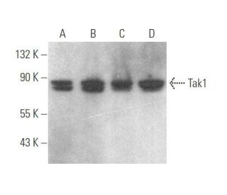
Date published: 2026-2-27

1-800-457-3801

SCBT Portrait Logo
Seach Input

Tak1 Antibody (H-5): sc-166562

5.0(5)
Write a reviewAsk a question

Datasheets
  • Tak1 Antibody (H-5) is a mouse monoclonal IgG3, cited in 19 publications, provided at 200 µg/ml
  • raised against a peptide mapping at the C-terminus of Tak1 of mouse origin
  • recommended for detection of Tak1 of mouse, rat and human origin by WB, IP, IF, IHC(P) and ELISA; also reactive with additional species, including and equine, bovine and porcine
  • See Tak1 (C-9): sc-7967 for Tak1 antibody conjugates, including AC, HRP, FITC, PE, Alexa Fluor® 488, 594, 647, 680 and 790.
  • m-IgGκ BP-HRP is the preferred secondary detection reagent for Tak1 Antibody (H-5) for WB and IHC(P) applications. This reagent is now offered in a bundle with Tak1 Antibody (H-5) (see ordering information below). For additional m-IgGκ BP conjugates see our complete list of Mouse IgG Binding Proteins.
Crispr Promo Banner

QUICK LINKS

SEE ALSO...

Tak1 Antibody (H-5) is a mouse monoclonal IgG3 antibody raised against a peptide mapping at the C-terminus of Tak1 (Transforming Growth Factor-beta Activated Kinase 1) of mouse origin, also known as MAP3K7 (Mitogen-Activated Protein Kinase Kinase Kinase 7). Tak1 (clone H-5) antibody detects Tak1 protein from mouse, rat, human, equine, bovine, and porcine sources by western blotting (WB), immunoprecipitation (IP), immunofluorescence (IF), immunohistochemistry with paraffin-embedded sections (IHCP), and enzyme-linked immunosorbent assay (ELISA). Tak1 is a critical serine/threonine kinase located primarily in the cytoplasm, where Tak1 plays a pivotal role in signal transduction pathways by mediating cellular responses to cytokines such as TGFβ (Transforming Growth Factor-beta) and interleukin-1 (IL-1). The cytoplasmic localization of Tak1 is essential for interacting with upstream activators and downstream effectors, influencing processes like inflammation, immune responses, cell survival, and apoptosis. Dysregulation of Tak1 function has been linked to various pathological conditions, including cancer and inflammatory diseases, making Tak1 an important target for biomedical research. Anti-Tak1 antibody (H-5) facilitates the study of key signaling pathways and their roles in disease mechanisms.

For Research Use Only. Not Intended for Diagnostic or Therapeutic Use.

Ordering Information

Product NameCatalog #UNITPriceQtyFAVORITES

Tak1 Antibody (H-5)

sc-166562
200 µg/ml
$322.00

Tak1 Antibody (H-5): m-IgGκ BP-HRP Bundle

sc-551982
200 µg Ab; 40 µg BP
$361.00

Tak1 (H-5) Neutralizing Peptide

sc-166562 P
100 µg/0.5 ml
$69.00

What is the recommended secondary reagent to be used with this mouse monoclonal primary?

Asked by: jerojero
We recommend using one our exclusive Mouse IgG Binding Proteins as a secondary detection reagent. A complete list of available binding proteins is available on our website here: https://www.scbt.com/scbt/browse/support-products-mouse-igg-binding-proteins/_/N-ecrety
Answered by: Tech Service 3
Date published: 2017-02-02
  • y_2026, m_2, d_25, h_10CST
  • bvseo_bulk, prod_bvqa, vn_bulk_3.0.42
  • cp_1, bvpage1
  • co_hasquestionsanswers, tq_1
  • loc_en_US, sid_166562, prod, sort_[SortEntry(order=LAST_APPROVED_ANSWER_SUBMISSION_TIME, direction=DESCENDING)]
  • clientName_scbt
  • bvseo_sdk, java_sdk, bvseo-4.0.0
  • CLOUD, getContent, 138ms
  • QUESTIONS, PRODUCT
Rated 5 out of 5 by from Good for IHCwe had good results using this antibody for IHC in mice pituitary slices, with sc-516102 as secondary
Date published: 2021-02-11
Rated 5 out of 5 by from Works well by immunostainingWorks well on mouse brain for immunostaining, with no background.
Date published: 2017-12-18
Rated 5 out of 5 by from Good western blotWe use this antibody for western blot and we got a good band around the correct molecular weight when using HeLa cells
Date published: 2017-01-11
Rated 5 out of 5 by from excellent!i used this antibody to detect mice TAK1 in spinal cord for Western Blot. it worked very well.
Date published: 2016-11-15
Rated 5 out of 5 by from Produced nice Western blot data of Tak1 expressionProduced nice Western blot data of Tak1 expression in 3611-RF whole cell lysate. -SCBT QC
Date published: 2014-10-11
  • y_2026, m_2, d_25, h_10
  • bvseo_bulk, prod_bvrr, vn_bulk_3.0.42
  • cp_1, bvpage1
  • co_hasreviews, tv_0, tr_5
  • loc_en_US, sid_166562, prod, sort_[SortEntry(order=SUBMISSION_TIME, direction=DESCENDING)]
  • clientName_scbt
  • bvseo_sdk, java_sdk, bvseo-4.0.0
  • CLOUD, getReviews, 15ms
  • REVIEWS, PRODUCT
Tak1 Antibody (H-5) is rated 5.0 out of 5 by 5.
  • y_2026, m_2, d_25, h_10
  • bvseo_bulk, prod_bvrr, vn_bulk_3.0.42
  • cp_1, bvpage1
  • co_hasreviews, tv_0, tr_5
  • loc_en_US, sid_166562, prod, sort_[SortEntry(order=SUBMISSION_TIME, direction=DESCENDING)]
  • clientName_scbt
  • bvseo_sdk, java_sdk, bvseo-4.0.0
  • CLOUD, getAggregateRating, 101ms
  • REVIEWS, PRODUCT