")
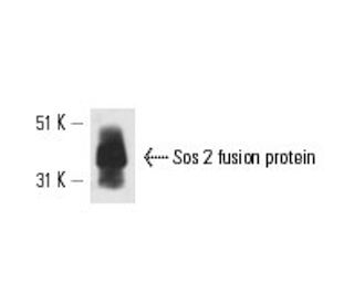
: sc-55478. Western blot analysis of human recombinant Sos 2 fusion protein.")
Sos 2 Antibody (A-8): sc-55478
- Sos 2 Antibody (A-8) is a mouse monoclonal IgG2a κ provided at 200 µg/ml
- raised against amino acids 1091-1170 mapping near the C-terminus of Sos 2 of human origin
- recommended for detection of Sos 2 of human origin by WB, IP, IF and ELISA
- m-IgG Fc BP-HRP is the preferred secondary detection reagent for Sos 2 Antibody (A-8) for WB applications. This reagent is now offered in a bundle with Sos 2 Antibody (A-8) (see ordering information below).

QUICK LINKS
Sos 2 Antibody (A-8) is a mouse monoclonal IgG2a antibody that detects Sos 2 in human samples through applications such as western blotting (WB), immunoprecipitation (IP), immunofluorescence (IF), and enzyme-linked immunosorbent assay (ELISA). Sos 2, a member of the guanine nucleotide exchange factor family, plays a crucial role in the activation of Ras proteins, which are pivotal in regulating various cellular processes, including growth, differentiation, and survival. Sos 2 is primarily located in the cytoplasm, where Sos 2 facilitates the exchange of GDP for GTP on Ras, thereby promoting its active form. This activation is essential for the downstream signaling pathways that lead to cellular responses, making anti-Sos 2 antibody (A-8) a key player in signal transduction. Furthermore, Sos 2 interacts with other proteins, such as receptor tyrosine kinases, which are vital for initiating the signaling cascade that leads to Ras activation. The ability of Sos 2 to modulate Ras activity underscores Sos 2′s importance in both normal cellular function and the pathogenesis of various diseases, including cancer, where aberrant Ras signaling is often observed. Anti-Sos 2 antibody (A-8) serves as an invaluable tool for researchers studying the intricate mechanisms of Ras signaling and its implications in health and disease.
Alexa Fluor® is a trademark of Molecular Probes Inc., OR., USA
LI-COR® and Odyssey® are registered trademarks of LI-COR Biosciences
Sos 2 Antibody (A-8) References:
- Candidate gene association study of insulin signaling genes and Alzheimer's disease: evidence for SOS2, PCK1, and PPARgamma as susceptibility loci. | Hamilton, G., et al. 2007. Am J Med Genet B Neuropsychiatr Genet. 144B: 508-16. PMID: 17440948
- Triggering Ras signalling by intracellular Francisella tularensis through recruitment of PKCα and βI to the SOS2/GrB2 complex is essential for bacterial proliferation in the cytosol. | Al-Khodor, S. and Abu Kwaik, Y. 2010. Cell Microbiol. 12: 1604-21. PMID: 20618341
- Rare variants in SOS2 and LZTR1 are associated with Noonan syndrome. | Yamamoto, GL., et al. 2015. J Med Genet. 52: 413-21. PMID: 25795793
- SOS2 and ACP1 Loci Identified through Large-Scale Exome Chip Analysis Regulate Kidney Development and Function. | Li, M., et al. 2017. J Am Soc Nephrol. 28: 981-994. PMID: 27920155
- SOS GEFs in health and disease. | Baltanás, FC., et al. 2020. Biochim Biophys Acta Rev Cancer. 1874: 188445. PMID: 33035641
- First prenatal case of Noonan syndrome with SOS2 mutation: Implications of early diagnosis for genetic counseling. | Gentile, M., et al. 2021. Am J Med Genet A. 185: 1897-1902. PMID: 33750022
- SOS2 Comes to the Fore: Differential Functionalities in Physiology and Pathology. | Baltanás, FC., et al. 2021. Int J Mol Sci. 22: PMID: 34205562
- Sulfarotene, a synthetic retinoid, overcomes stemness and sorafenib resistance of hepatocellular carcinoma via suppressing SOS2-RAS pathway. | Qi, F., et al. 2021. J Exp Clin Cancer Res. 40: 280. PMID: 34479623
- Lymphatic anomalies during lifetime in patients with Noonan syndrome: Retrospective cohort study. | Swarts, JW., et al. 2022. Am J Med Genet A. 188: 3242-3261. PMID: 35979676
- Chromosomal localization of two genes encoding human ras exchange factors: SOS1 maps to the 2p22-->p16 region and SOS2 to the 14q21-->q22 region of the human genome. | Chardin, P. and Mattei, MG. 1994. Cytogenet Cell Genet. 66: 68-9. PMID: 8275713
Ordering Information
| Product Name | Catalog # | UNIT | Price | Qty | FAVORITES | |
Sos 2 Antibody (A-8) | sc-55478 | 200 µg/ml | $322.00 | |||
Sos 2 Antibody (A-8): m-IgG Fc BP-HRP Bundle | sc-539020 | 200 µg Ab; 10 µg BP | $361.00 |