")
PC-1 Antikörper (H-7): sc-393419
- PC-1 Antikörper H-7 ist ein Maus monoklonales IgG2b κ PC-1 Antikörper, verwendet in 3 wissenschaftlichen Veröffentlichungen, in einer Menge von 200 µg/ml
- spezifisch für ein Epitop, welches zwischen den Aminosäuren 9-36 am N-terminus von PC-1 aus der Spezies human liegt
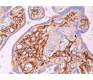
- PC-1 Antikörper (H-7) ist empfohlen für die Detektion von PC-1 aus der Spezies mouse, rat und human per WB, IP, IF, IHC(P) und ELISA
- Anti-PC-1 Antikörper (H-7) ist erhältlich als Konjugat mit Agarose für IP; HRP für WB, IHC(P) und ELISA; und entweder mit Phycoerythrin oder FITC für IF, IHC(P) und FCM
- auch erhältlich als Konjugat mit Alexa Fluor® 488, Alexa Fluor® 546, Alexa Fluor® 594 oder Alexa Fluor® 647 für IF, IHC(P) und FCM
- auch erhältlich als Konjugat mit Alexa Fluor® 680 oder Alexa Fluor® 790 für WB (NIR), IF und FCM
- m-IgG Fc BP-HRP, 2b BP-HRP">m-IgG2b BP-HRP und m-IgGκ BP-HRP sind die bevorzugten sekundären Nachweisreagenzien für PC-1 Antikörper (H-7) für WB- und IHC(P)-Anwendungen. Diese Reagenzien werden jetzt in Bündeln mit PC-1 Antikörper (H-7) angeboten(siehe Bestellinformationen unten).

Direktverknüpfungen
Siehe auch...
PC-1-Antikörper (H-7) ist ein monoklonaler IgG2b κ-Maus-Antikörper gegen PC-1 (auch als PC-1-Antikörper bezeichnet), der das PC-1-Protein von Maus, Ratte und Mensch mittels WB, IP, IF, IHC(P) und ELISA nachweist. Der PC-1-Antikörper (H-7) ist sowohl als nicht konjugierter Anti-PC-1-Antikörper als auch als mehrfach konjugierter Anti-PC-1-Antikörper erhältlich, darunter Agarose-, HRP-, PE-, FITC- und mehrere Alexa Fluor®-Konjugate. PC-1, auch bekannt als Ektonukleotid-Pyrophosphatase/Phosphodiesterase 1 (ENPP1) oder Membrankomponente, Chromosom 6, Oberflächenmarker-1 (M6S1), ist das menschliche Homolog von Ly-41 in der Maus. PC-1 ist ein Homodimer mit begrenzter Gewebeverteilung, das zuerst in Plasmazellen charakterisiert wurde. Neben seiner Expression auf Plasmazellen wird PC-1 auch auf Hepatozyten, Nierentubuli, Speicheldrüsenepithel, Epididymis, Kapillarendothel im Gehirn und Chondrozyten exprimiert. Die meisten Patienten mit nicht insulinabhängigem Diabetes mellitus (NIDDM) sind sowohl gegen endogenes als auch gegen exogenes Insulin resistent. Die Insulinresistenz geht dem Ausbruch dieser Krankheit voraus, was darauf hindeutet, dass es sich um eine anfängliche Anomalie handeln könnte. Es wurde vermutet, dass PC-1 durch direkte Interaktion mit der α-Untereinheit des Rezeptors eine Rolle bei der Insulinresistenz von NIDDM spielen könnte. Das Gen, das für PC-1 kodiert, ist auf dem menschlichen Chromosom 6q22-q23 kartiert, das eine häufige Stelle für Deletionen bei menschlichen lymphatischen Neoplasien ist.
Alexa Fluor® ist ein Markenzeichen von Molecular Probes Inc., OR., USA
LI-COR® und Odyssey® sind Markenzeichen von LI-COR Biosciences
PC-1 Antikörper (H-7) Literaturhinweise:
- Das Membranglykoprotein PC-1 hemmt die Funktion des Insulinrezeptors durch direkte Interaktion mit der Alpha-Untereinheit des Rezeptors. | Maddux, BA. and Goldfine, ID. 2000. Diabetes. 49: 13-9. PMID: 10615944
- Das Plasmazellmembran-Glykoprotein-Gen Pca-1 (alkalische Phosphodiesterase I) ist mit dem Proto-Onkogen Myb auf dem Maus-Chromosom 10 verbunden. | Buckley, MF. and Goding, JW. 1992. Immunogenetics. 36: 199-201. PMID: 1351877
- Plasmazellmembran-Glykoprotein PC-1. cDNA-Klonierung des menschlichen Moleküls, Aminosäuresequenz und chromosomale Lage. | Buckley, MF., et al. 1990. J Biol Chem. 265: 17506-11. PMID: 2211644
- Verteilung des murinen Plasmazellantigens PC-1 in nicht-lymphoiden Geweben. | Harahap, AR. and Goding, JW. 1988. J Immunol. 141: 2317-20. PMID: 3262656
- Oberflächenalloantigene von Plasmazellen. | Takahashi, T., et al. 1970. J Exp Med. 131: 1325-41. PMID: 5419273
Bestellinformation
| Produkt | Katalog # | EINHEIT | Preis | ANZAHL | Favoriten | |
PC-1 Antikörper (H-7) | sc-393419 | 200 µg/ml | $322.00 | |||
PC-1 (H-7): m-IgG Fc BP-HRP Bundle | sc-530450 | 200 µg Ab; 10 µg BP | $361.00 | |||
PC-1 (H-7): m-IgGκ BP-HRP Bundle | sc-523938 | 200 µg Ab, 40 µg BP | $361.00 | |||
PC-1 (H-7): m-IgG2b BP-HRP Bundle | sc-550189 | 200 µg Ab; 10 µg BP | $361.00 | |||
PC-1 Antikörper (H-7) AC | sc-393419 AC | 500 µg/ml, 25% agarose | $424.00 | |||
PC-1 Antikörper (H-7) HRP | sc-393419 HRP | 200 µg/ml | $322.00 | |||
PC-1 Antikörper (H-7) FITC | sc-393419 FITC | 200 µg/ml | $336.00 | |||
PC-1 Antikörper (H-7) PE | sc-393419 PE | 200 µg/ml | $349.00 | |||
PC-1 Antikörper (H-7) Alexa Fluor® 488 | sc-393419 AF488 | 200 µg/ml | $364.00 | |||
PC-1 Antikörper (H-7) Alexa Fluor® 546 | sc-393419 AF546 | 200 µg/ml | $364.00 | |||
PC-1 Antikörper (H-7) Alexa Fluor® 594 | sc-393419 AF594 | 200 µg/ml | $364.00 | |||
PC-1 Antikörper (H-7) Alexa Fluor® 647 | sc-393419 AF647 | 200 µg/ml | $364.00 | |||
PC-1 Antikörper (H-7) Alexa Fluor® 680 | sc-393419 AF680 | 200 µg/ml | $364.00 | |||
PC-1 Antikörper (H-7) Alexa Fluor® 790 | sc-393419 AF790 | 200 µg/ml | $364.00 | |||
PC-1 (H-7) Neutralizing Peptid | sc-393419 P | 100 µg/0.5 ml | $69.00 |