
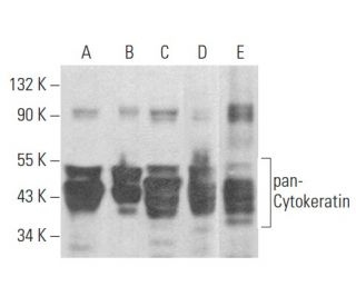
pan-Cytokeratin Antibody (B311.1): sc-58826
- pan-Cytokeratin Antibody (B311.1) is a mouse monoclonal IgG1 κ, cited in 5 publications, provided at 200 µg/ml
- raised against a pan-Cytokeratin fraction isolated from A-431 epidermoid carcinoma cell line of human origin
- recommended for detection of Cytokeratin 4, 5, 6, 8, 10, 13 and 18 of mouse, rat, human and bovine origin by WB, IP, IF, IHC(P), FCM and ELISA
- See pan-Cytokeratin (C11): sc-8018 for pan-Cytokeratin antibody conjugates, including AC, HRP, FITC, PE, Alexa Fluor® 488, 594, 647, 680 and 790.
- m-IgG Fc BP-HRP, m-IgG1 BP-HRP and m-IgGκ BP-HRP are the preferred secondary detection reagents for pan-Cytokeratin Antibody (B311.1) for WB and IHC(P) applications. These reagents are now offered in bundles with pan-Cytokeratin Antibody (B311.1) (see ordering information below).

QUICK LINKS
SEE ALSO...
pan-Cytokeratin Antibody (B311.1) is a mouse monoclonal IgG1 antibody that detects pan-Cytokeratin in mouse, rat, human, and bovine samples through various applications including western blotting (WB), immunoprecipitation (IP), immunofluorescence (IF), immunohistochemistry with paraffin-embedded sections (IHCP), flow cytometry (FCM), and enzyme-linked immunosorbent assay (ELISA). pan-Cytokeratin is a crucial component of the cytoskeletal structure in epithelial cells, providing mechanical support and playing a vital role in maintaining cell shape and integrity. pan-Cytokeratin is primarily located in the cytoplasm of epithelial cells, where pan-Cytokeratin forms a network of intermediate filaments that contribute to the overall architecture of tissues. This structural role is particularly important in the context of tissue resilience and protection against mechanical stress, which is essential for the proper functioning of epithelial tissues. Additionally, pan-Cytokeratin serves as a valuable biomarker for identifying and characterizing various types of epithelial tumors, as pan-Cytokeratin expression patterns can indicate the differentiation status of malignant cells. For instance, specific cytokeratin isoforms are often upregulated in certain carcinomas, making pan-Cytokeratin (B311.1) antibody a key tool in cancer diagnostics and research.
Alexa Fluor® is a trademark of Molecular Probes Inc., OR., USA
LI-COR® and Odyssey® are registered trademarks of LI-COR Biosciences
pan-Cytokeratin Antibody (B311.1) References:
- The usefulness of cytokeratin immunohistochemistry in detection of lymph node micrometastasis in neck dissection specimens. | Kwon, SY., et al. 2004. Otolaryngol Head Neck Surg. 131: 300-6. PMID: 15365551
- Distinction of high-grade neuroendocrine carcinoma/small cell carcinoma from conventional urothelial carcinoma of urinary bladder: an immunohistochemical approach. | Thompson, S., et al. 2011. Appl Immunohistochem Mol Morphol. 19: 395-9. PMID: 21617524
- Characterization of LHY-821, a novel moderately differentiated endometrial carcinoma cell line. | Hu, Q., et al. 2012. In Vitro Cell Dev Biol Anim. 48: 441-8. PMID: 22806968
- Immunohistochemical expression of cytokeratins and epithelial membrane protein 2 in nasopharyngeal carcinoma and its potential implications. | Ahmed, HG., et al. 2015. Asian Pac J Cancer Prev. 16: 653-6. PMID: 25684502
- Diverse Immunoprofile of Ductal Adenocarcinoma of the Prostate with an Emphasis on the Prognostic Factors. | Jeong, SU., et al. 2017. J Pathol Transl Med. 51: 471-481. PMID: 28793393
- The Pan-Cytokeratin Staining Intensity and Fractal Computational Analysis of Breast Tumor Malignant Growth Patterns Prognosticate the Occurrence of Distant Metastasis. | Rajković, N., et al. 2018. Front Oncol. 8: 348. PMID: 30214894
- Histopathological Significance and Prognostic Impact of Tumor Budding in Colorectal Cancer. | Mehta, A., et al. 2018. Asian Pac J Cancer Prev. 19: 2447-2453. PMID: 30255698
- Three Dimensional Culture of Potential Epithelial Progenitor Cells in Human Lacrimal Gland. | Lin, H., et al. 2019. Transl Vis Sci Technol. 8: 32. PMID: 31523489
- Primary Ewing's sarcoma of the lung: a rare case report. | Anne, S., et al. 2020. Indian J Thorac Cardiovasc Surg. 36: 409-411. PMID: 33061150
- Spontaneous mandibular follicular ameloblastoma in a female Sprague-Dawley rat. | Li, J., et al. 2024. J Toxicol Pathol. 37: 39-43. PMID: 38283374
Ordering Information
| Product Name | Catalog # | UNIT | Price | Qty | FAVORITES | |
pan-Cytokeratin Antibody (B311.1) | sc-58826 | 200 µg/ml | $322.00 | |||
pan-Cytokeratin Antibody (B311.1): m-IgG Fc BP-HRP Bundle | sc-537156 | 200 µg Ab; 10 µg BP | $361.00 | |||
pan-Cytokeratin Antibody (B311.1): m-IgGκ BP-HRP Bundle | sc-534352 | 200 µg Ab; 40 µg BP | $361.00 | |||
pan-Cytokeratin Antibody (B311.1): m-IgG1 BP-HRP Bundle | sc-545041 | 200 µg Ab; 20 µg BP | $361.00 |