")
Anticorps pan-Cytokeratin (C11): sc-8018
- L'anticorps pan-Cytokeratin C11 est un monoclonal IgG1 κ L'Anticorps pan-Cytokeratin, cité dans 164 publications, fourni en 200 µg/ml
- élevé contre la lignée cellulaire de carcinome épidermoïde A-431 enrichie en kératine human.
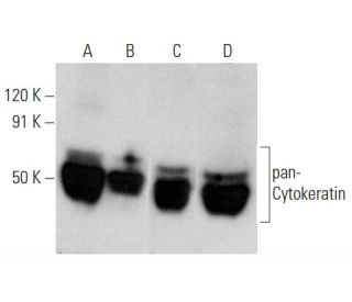
- L'Anticorps pan-Cytokeratin (C11) est recommandé pour la détection de Cytokeratin 4, 5, 6, 8, 10, 13 and 18 d'origine mouse, rat et human par WB, IP, IF, IHC(P), FCM et ELISA
- Anti-L'Anticorps pan-Cytokeratin (C11) est disponible conjugué à l'agarose pour IP; à l'HRP pour WB, IHC(P) et ELISA; et soit à la phycoerythrin ou FITC pour IF, IHC(P) et FCM
- aussi disponible conjugué à l'Alexa Fluor® 488, Alexa Fluor® 546, Alexa Fluor® 594 ou Alexa Fluor® 647 pour WB (RGB), IF, IHC(P) et FCM
- aussi disponible conjugué à l'Alexa Fluor® 680 ou Alexa Fluor® 790 pour WB (NIR), IF et FCM
- m-IgG Fc BP-HRP, 1 BP-HRP">m-IgG1 BP-HRP et m-IgGκ BP-HRP sont les réactifs de détection secondaire préférés pour l'anticorps pan-Cytokeratin (C11) pour les applications WB et IHC(P). Ces réactifs sont désormais proposés en lots avec l'anticorps pan-Cytokeratin (C11)(voir les informations de commande ci-dessous).

ACCÈS RAPIDE AUX LIENS
VOIR ÉGALEMENT...
L'anticorps pan-cytokératine (C11) est un anticorps monoclonal IgG1 de chaîne légère kappa de souris qui détecte la protéine pan-cytokératine d'origine humaine, murine et rat par western blotting (WB), immunoprécipitation (IP), immunofluorescence (IF), immunohistochimie sur coupes incluses en paraffine (IHCP), cytométrie de flux (FCM) et dosage immunoenzymatique (ELISA). L'anticorps pan-cytokératine (C11) est disponible sous forme non conjuguée et sous diverses formes conjuguées, notamment agarose, peroxydase de raifort (HRP), phycoérythrine (PE), isothiocyanate de fluorescéine (FITC) et plusieurs conjugués Alexa Fluor®. Les cytokératines, qui constituent un groupe diversifié de protéines de filaments intermédiaires, sont essentielles au maintien de l'intégrité structurelle des cellules épithéliales et sont exprimées par paires dans les tissus épithéliaux kératinisés et non kératinisés. L'importance des cytokératines s'étend à leur rôle dans la différenciation cellulaire et la spécialisation des tissus, ce qui fait de ces protéines des marqueurs précieux pour l'identification et la caractérisation des tumeurs malignes. Par exemple, des cytokératines spécifiques telles que la cytokératine 10 et la cytokératine 13 sont fortement exprimées dans certains carcinomes épidermoïdes, tandis que la cytokératine 18 se trouve principalement dans les adénocarcinomes et les carcinomes basocellulaires. La reconnaissance de ces protéines par l'anticorps anti-pan-Cytokératine (C11) renforce l'utilité de la recherche dans la recherche et le diagnostic du cancer, en fournissant des informations sur la biologie des tumeurs et en aidant à l'évaluation de la différenciation des tissus.
Alexa Fluor® est une marque déposée de Molecular Probes Inc., OR., USA
LI-COR® et Odyssey® sont marques déposées de LI-COR Biosciences
Anticorps pan-Cytokeratin (C11) Références:
- L'expression de K15 implique une différenciation latérale au sein des cellules basales épithéliales stratifiées. | Porter, RM., et al. 2000. Lab Invest. 80: 1701-10. PMID: 11092530
- Étude de l'expression de la cytokératine dans les kystes épithéliaux de la peau et dans certains types peu courants de tumeurs kystiques, à l'aide d'anticorps spécifiques à la chaîne. | Broekaert, D., et al. 1990. Arch Dermatol Res. 282: 383-91. PMID: 1701984
- Tumeurs pulmonaires humaines: corrélation entre le profil antigénique et le type histologique. | Gatter, KC., et al. 1985. Histopathology. 9: 805-23. PMID: 2414203
- Caractérisation de deux anticorps monoclonaux anti-kératine et leur utilisation dans l'étude des troubles épithéliaux. | Pulford, KA., et al. 1985. Histopathology. 9: 825-40. PMID: 2414204
- Marqueurs de la pan-cytokératine pour l'immunocytochimie rapide de coupes congelées de cas de Mohs de carcinome basocellulaire de la tête et du visage: une comparaison et une évaluation pour déterminer le marqueur de choix. | Orchard, GE., et al. 2015. Br J Biomed Sci. 72: 61-6. PMID: 26126321
- [Évaluation de l'immunohistochimie de la pan-cytokératine et de la double coloration au bleu Victoria dans l'évaluation de la stadification du cancer colorectal pT3 et pT4a]. | Xie, LW., et al. 2023. Zhonghua Bing Li Xue Za Zhi. 52: 1034-1036. PMID: 37805397
- Évaluation d'un nouveau marqueur tumoral pour les fragments de cytokératine 8 et 18 chez des individus sains et des patients atteints de cancer de la prostate. | Silén, A., et al. 1994. Prostate. 24: 326-32. PMID: 7516071
- Un nouvel IRMA et ELISA pour quantifier les fragments de cytokératine 8 et 18 dans le sérum d'individus sains et de patients atteints de cancer. | Silén, A., et al. 1995. Scand J Clin Lab Invest. 55: 153-61. PMID: 7545308
- Expression de la cytokératine dans les épithéliums normaux et (pré)malins de la tête et du cou: une vue d'ensemble. | van der Velden, LA., et al. 1993. Head Neck. 15: 133-46. PMID: 7680025
- Distribution sérique et tissulaire d'un fragment de la cytokératine 19 (cyfra 21-1) chez des patients atteints de cancer du poumon. | Quillien, V., et al. 1995. Anticancer Res. 15: 2857-63. PMID: 8669879
- L'inactivation fonctionnelle de p53 par l'ARN antisens induit la capacité invasive des cellules de carcinome pulmonaire et régule la synthèse de la cytokératine. | Mukhopadhyay, T. and Roth, JA. 1996. Anticancer Res. 16: 1683-9. PMID: 8712687
- Expression de la cytokératine, organisation fibrillaire et fonction subtile dans les cellules hépatiques. | Marceau, N. and Loranger, A. 1995. Biochem Cell Biol. 73: 619-25. PMID: 8714681
Informations pour la commande
| Nom du produit | Ref. Catalogue | COND. | Prix HT | QTÉ | Favoris | |
Anticorps pan-Cytokeratin (C11) | sc-8018 | 200 µg/ml | $322.00 | |||
pan-Cytokeratin (C11): m-IgG Fc BP-HRP Kit | sc-528202 | 200 µg Ab; 10 µg BP | $361.00 | |||
pan-Cytokeratin (C11): m-IgGκ BP-HRP Kit | sc-520524 | 200 µg Ab, 40 µg BP | $361.00 | |||
pan-Cytokeratin (C11): m-IgG1 BP-HRP Kit | sc-542813 | 200 µg Ab; 20 µg BP | $361.00 | |||
Anticorps pan-Cytokeratin (C11) AC | sc-8018 AC | 500 µg/ml, 25% agarose | $424.00 | |||
Anticorps pan-Cytokeratin (C11) HRP | sc-8018 HRP | 200 µg/ml | $322.00 | |||
Anticorps pan-Cytokeratin (C11) FITC | sc-8018 FITC | 200 µg/ml | $336.00 | |||
Anticorps pan-Cytokeratin (C11) PE | sc-8018 PE | 200 µg/ml | $349.00 | |||
Anticorps pan-Cytokeratin (C11) Alexa Fluor® 488 | sc-8018 AF488 | 200 µg/ml | $364.00 | |||
Anticorps pan-Cytokeratin (C11) Alexa Fluor® 546 | sc-8018 AF546 | 200 µg/ml | $364.00 | |||
Anticorps pan-Cytokeratin (C11) Alexa Fluor® 594 | sc-8018 AF594 | 200 µg/ml | $364.00 | |||
Anticorps pan-Cytokeratin (C11) Alexa Fluor® 647 | sc-8018 AF647 | 200 µg/ml | $364.00 | |||
Anticorps pan-Cytokeratin (C11) Alexa Fluor® 680 | sc-8018 AF680 | 200 µg/ml | $364.00 | |||
Anticorps pan-Cytokeratin (C11) Alexa Fluor® 790 | sc-8018 AF790 | 200 µg/ml | $364.00 |