")
, 钙culin A 处理 (B, E) 和钙culin A 及λ蛋白磷酸酶 (sc-200312A) 处理 (C, F) 的 Jurkat 全细胞裂解液中 Akt1 磷酸化的 Western 印迹分析. 测试的抗体包括 p-Akt1 抗体 (5. Ser 473): sc-293125 (A, B, C) 和 Akt1 (C-20): sc-1618 (D, E, F).")
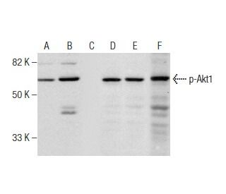
, 经 mLIF 处理 (B, E) 以及经 mLIF 和 lambda 蛋白磷酸酶 (sc-200312A) 处理 (C, F) 的 3T3-L1 全细胞裂解液中 Akt1 磷酸化的 Western 印迹分析. 测试的抗体包括 p-Akt1 抗体 (5. Ser 473): sc-293125 (A, B, C) 和 Akt1 (C-20): sc-1618 (D, E, F).")
 HRP: sc-293125 HRP. Jurkat (A) 和 Jurkat + PMA 诱导 (B) 全细胞裂解液中 Akt1 磷酸化的直接 Western 印迹分析.")
未经处理 (A, D), 钙culin A 处理 (B, E) 和钙culin A 及λ蛋白磷酸酶 (sc-200312A) 处理 (C, F) 的 Jurkat 全细胞裂解液中 Akt1 磷酸化的 Western 印迹分析. 测试的抗体包括 p-Akt1 抗体 (5. Ser 473): sc-293125 (A, B, C) 和 Akt1 (C-20): sc-1618 (D, E, F).
p-Akt1 抗体 (5.Ser 473): sc-293125
- p-Akt1抗体(5.Ser 473)是小鼠单克隆IgG1 κ, 在129篇文献中引用,规格为200 µg/ml
- 针对含磷酸化Ser 473的短氨基酸序列的抗体,该序列来自Akt1 of human
- 推荐用于 mouse, rat 和 human 来源的Ser 473 phosphorylated Akt1 WB, IP 和 ELISA检测; 可能与 Ser 473 phosphorylated Akt2 and Ser 473 phosphorylated Akt3产生交联反应
- 还可偶联琼脂糖 用于IP; 偶联HRP 用于WB, IHC(P) 和ELISA
- m-IgG Fc BP-HRP、 1 BP-HRP">m-IgG1 BP-HRP和m-IgGκ BP-HRP是p-Akt1 Antibody (5.Ser 473) 适用于 WB 应用。 的首选辅助检测试剂。这些试剂现与p-Akt1 Antibody (5.Ser 473) 打包提供(请参阅下面的订购信息)。

p-Akt1抗体(5.Ser 473)是一种IgG1 κ小鼠单克隆p-Akt1抗体,可通过WB、IP和ELISA检测小鼠、大鼠和人类来源的Ser 473磷酸化Akt1。p-Akt1抗体(5.Ser 473)可作为偶联和非偶联的抗p-Akt1抗体形式提供。丝氨酸/苏氨酸激酶Akt家族包含几个成员,包括Akt1(也称为PKB或RacPK)、Akt2(也称为PKBβ或RacPK-β)和Akt 3(也称为PKBγ或thyoma病毒原发癌基因3),它们与蛋白激酶A和C家族具有序列同源性,并由c-Akt原癌基因编码。Akt家族的所有成员都具有血小板同源结构域。Akt1和Akt2通过PDGF刺激被激活。这种激活依赖于PDGFR-β酪氨酸残基740和751,它们与磷脂酰肌醇3-激酶(PI 3-激酶)复合物的亚基结合。胰岛素或胰岛素生长因子-1(IGF-1)对Akt1的激活导致Thr 308和Ser 473的磷酸化。在胰岛素/IGF-1刺激的细胞中,Akt蛋白通过上游激酶被磷酸化和激活,而Akt1和Akt2的激活被PI激酶抑制剂wortmannin抑制。综上所述,这些数据充分表明该蛋白是PI激酶下游的信号分子。Akt3在丝氨酸残基上被磷酸化以响应胰岛素。然而,胰岛素对Akt3的激活被蛋白激酶C的先前激活所抑制,这种机制不需要PH结构域的存在。Akt3在3T3-L1成纤维细胞、脂肪细胞和骨骼肌中表达,可能参与各种生物过程,包括脂肪细胞和肌肉分化、糖原合成、葡萄糖摄取、细胞凋亡和细胞增殖。
仅限研究使用。不适用于诊断和治疗用途。
Alexa Fluor® 是Molecular Probes Inc., OR., USA的商标
LI-COR®和 Odyssey® 是LI-COR Biosciences的注册商标
p-Akt1 抗体 (5.Ser 473) 参考文献:
- 鉴定含有丝氨酸磷酸化调节位点的人类 Akt3(蛋白激酶 B γ)。 | Nakatani, K., et al. 1999. Biochem Biophys Res Commun. 257: 906-10. PMID: 10208883
- 通过荧光原位杂交将编码Akt/蛋白激酶B家族成员的AKT3绘制到人类和啮齿动物的染色体上。 | Murthy, SS., et al. 2000. Cytogenet Cell Genet. 88: 38-40. PMID: 10773662
- 与胃癌细胞周期异常相关的新型 AKT1 突变。 | Ghatak, S., et al. 2018. Per Med. 15: 79-86. PMID: 29714127
- 外周支气管乳头状瘤中的 AKT1 基因突变 腺乳头状瘤和鳞状细胞与腺乳头状瘤混合瘤与支气管腺瘤的区别。 | Sasaki, E., et al. 2021. Am J Surg Pathol. 45: 119-126. PMID: 32868527
- 多发性硬化症患者外周T细胞亚群中Akt1和p-Akt1的表达。 | Oktelik, FB., et al. 2021. Acta Neurol Belg. 121: 1777-1782. PMID: 33034831
- AKT1(p. E17K)热点突变对恶性肿瘤发生和预后的影响 | Chen, Y., et al. 2020. Front Cell Dev Biol. 8: 573599. PMID: 33123537
- 磷脂酰肌醇-3-OH 激酶信号转导中的蛋白激酶 B(c-Akt)。 | Burgering, BM. and Coffer, PJ. 1995. Nature. 376: 599-602. PMID: 7637810
- Akt 原癌基因编码的蛋白激酶是 PDGF 激活的磷脂酰肌醇 3- 激酶的靶标。 | Franke, TF., et al. 1995. Cell. 81: 727-36. PMID: 7774014
- AH/PH 结构域介导的 Akt 分子间相互作用及其在 Akt 调节中的潜在作用。 | Datta, K., et al. 1995. Mol Cell Biol. 15: 2304-10. PMID: 7891724
- 人胰腺细胞中 AKT2 的扩增以及反义 RNA 对 AKT2 表达和致瘤性的抑制。 | Cheng, JQ., et al. 1996. Proc Natl Acad Sci U S A. 93: 3636-41. PMID: 8622988
- 蛋白激酶 C 可调节 3T3-L1 脂肪细胞在胰岛素刺激下增加的 Akt1 和 Akt3 活性。 | Barthel, A., et al. 1998. Biochem Biophys Res Commun. 243: 509-13. PMID: 9480839
- 体内大鼠骨骼肌中的 Akt 激酶和 2-脱氧葡萄糖摄取:与胰岛素和运动有关的研究。 | Turinsky, J. and Damrau-Abney, A. 1999. Am J Physiol. 276: R277-82. PMID: 9887206
订购信息
| 产品名称 | 产品编号 | 规格 | 价格 | 数量 | 收藏夹 | |
p-Akt1 抗体 (5.Ser 473) | sc-293125 | 200 µg/ml | $322.00 | |||
p-Akt1 (5.Ser 473): m-IgG Fc BP-HRP 套装 | sc-529297 | 200 µg Ab; 10 µg BP | $361.00 | |||
p-Akt1 (5.Ser 473): m-IgGκ BP-HRP 套装 | sc-522114 | 200 µg Ab, 40 µg BP | $361.00 | |||
p-Akt1 (5.Ser 473): m-IgG1 BP-HRP 套装 | sc-543437 | 200 µg Ab; 20 µg BP | $361.00 | |||
p-Akt1 抗体 (5.Ser 473) AC | sc-293125 AC | 500 µg/ml, 25% agarose | $424.00 | |||
p-Akt1 抗体 (5.Ser 473) HRP | sc-293125 HRP | 200 µg/ml | $322.00 |