Date published: 2025-12-7

1-800-457-3801

SCBT Portrait Logo
Seach Input

Occludin Antibody (E-5): sc-133256

4.2(37)
Write a reviewAsk a question

Datasheets
  • Occludin Antibody (E-5) is a mouse monoclonal IgG2b κ Occludin antibody, cited in 161 publications, provided at 200 µg/ml
  • raised against amino acids 132-411 mapping within an internal region of Occludin of human origin
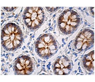
  • Occludin Antibody (E-5) is recommended for detection of Occludin of mouse, rat and human origin by WB, IP, IF, IHC(P) and ELISA
  • Anti-Occludin Antibody (E-5) is available conjugated to agarose for IP; HRP for WB, IHC(P) and ELISA; and to either phycoerythrin or FITC for IF, IHC(P) and FCM
  • also available conjugated to Alexa Fluor® 488, Alexa Fluor® 546, Alexa Fluor® 594 or Alexa Fluor® 647 for WB (RGB), IF, IHC(P) and FCM, and for use with RGB fluorescent imaging systems, such as iBright™ FL1000, FluorChem™, Typhoon, Azure and other comparable systems
  • also available conjugated to Alexa Fluor® 680 or Alexa Fluor® 790 for WB (NIR), IF and FCM; for use with Near-Infrared (NIR) detection systems, such as LI-COR®Odyssey®, iBright™ FL1000, FluorChem™, Typhoon, Azure and other comparable systems
  • m-IgG2b BP-HRP and m-IgGκ BP-HRP are the preferred secondary detection reagents for Occludin Antibody (E-5) for WB and IHC(P) applications. These reagents are now offered in bundles with Occludin Antibody (E-5) (see ordering information below).

QUICK LINKS

SEE ALSO...

Occludin Antibody (E-5) is a mouse monoclonal IgG2b kappa light chain antibody that detects Occludin protein of mouse, rat, and human origin by western blotting (WB), immunoprecipitation (IP), immunofluorescence (IF), immunohistochemistry with paraffin-embedded sections (IHCP), and enzyme-linked immunosorbent assay (ELISA). Anti-Occludin antibody (E-5) is available in both non-conjugated and various conjugated forms, including agarose, horseradish peroxidase (HRP), phycoerythrin (PE), fluorescein isothiocyanate (FITC), and multiple Alexa Fluor® conjugates. Occludin is an integral membrane protein that plays a crucial role in forming and maintaining tight junctions in epithelial and endothelial cells, which are essential for regulating paracellular permeability and maintaining blood-brain barrier integrity. Occludin features a tetraspan structure with two extracellular loops that facilitate interactions with other tight junction proteins, contributing to the overall architecture and function of the tight junction complex. Post-translational modifications, including phosphorylation, are vital for Occludin localization and functional regulation. Phosphorylation of Occludin can influence tight junction formation, impacting cellular permeability to large molecules. Polyunsaturated fatty acids up-regulate Occludin expression, which enhances transendothelial cell resistance, further highlighting Occludin′s importance in maintaining cellular barriers. Occludin shows high expression at cell-cell contact sites in brain tissue, while non-neural tissues display lower expression levels and a more discontinuous distribution. Changes in Occludin expression may enhance paracellular permeability and could be linked to tight junction damage in various pathological conditions.

For Research Use Only. Not Intended for Diagnostic or Therapeutic Use.

Alexa Fluor® is a trademark of Molecular Probes Inc., OR., USA

LI-COR® and Odyssey® are registered trademarks of LI-COR Biosciences

Occludin Antibody (E-5) References:

  1. Occludin degradation makes brain microvascular endothelial cells more vulnerable to reperfusion injury in vitro.  |  Zhang, Y., et al. 2021. J Neurochem. 156: 352-366. PMID: 32531803
  2. Occludin Promotes Adhesion of CD8+ T Cells and Melanocytes in Vitiligo via the HIF-1α Signaling Pathway.  |  Zou, P., et al. 2022. Oxid Med Cell Longev. 2022: 6732972. PMID: 35222802
  3. Higher serum occludin after successful reperfusion Is associated with early neurological deterioration.  |  Li, W., et al. 2022. CNS Neurosci Ther. 28: 999-1007. PMID: 35338575
  4. Cleavage of Occludin by Cigarette Smoke-Elicited Cathepsin S Increases Permeability of Lung Epithelial Cells.  |  Bigot, P., et al. 2022. Antioxidants (Basel). 12: PMID: 36670867
  5. The tight junction protein occludin modulates blood-brain barrier integrity and neurological function after ischemic stroke in mice.  |  Sugiyama, S., et al. 2023. Sci Rep. 13: 2892. PMID: 36806348
  6. Roles of epidermal growth factor receptor, claudin-1 and occludin in multi-step entry of hepatitis C virus into polarized hepatoma spheroids.  |  So, CW., et al. 2023. PLoS Pathog. 19: e1011887. PMID: 38157366
  7. Occludin: a novel integral membrane protein localizing at tight junctions.  |  Furuse, M., et al. 1993. J Cell Biol. 123: 1777-88. PMID: 8276896
  8. Molecular dissection of tight junctions.  |  Tsukita, S., et al. 1996. Cell Struct Funct. 21: 381-5. PMID: 9118244
  9. Possible involvement of phosphorylation of occludin in tight junction formation.  |  Sakakibara, A., et al. 1997. J Cell Biol. 137: 1393-401. PMID: 9182670
  10. Occludin as a possible determinant of tight junction permeability in endothelial cells.  |  Hirase, T., et al. 1997. J Cell Sci. 110 (Pt 14): 1603-13. PMID: 9247194
  11. Phosphorylation of occludin correlates with occludin localization and function at the tight junction.  |  Wong, V. 1997. Am J Physiol. 273: C1859-67. PMID: 9435490
  12. Regulation of tight junction permeability and occludin expression by polyunsaturated fatty acids.  |  Jiang, WG., et al. 1998. Biochem Biophys Res Commun. 244: 414-20. PMID: 9514943

Ordering Information

Product NameCatalog #UNITPriceQtyFAVORITES

Occludin Antibody (E-5)

sc-133256
200 µg/ml
$316.00

Occludin Antibody (E-5): m-IgGκ BP-HRP Bundle

sc-521292
200 µg Ab, 40 µg BP
$354.00

Occludin Antibody (E-5): m-IgG2b BP-HRP Bundle

sc-548883
200 µg Ab; 10 µg BP
$354.00

Occludin Antibody (E-5) AC

sc-133256 AC
500 µg/ml, 25% agarose
$416.00

Occludin Antibody (E-5) HRP

sc-133256 HRP
200 µg/ml
$316.00

Occludin Antibody (E-5) FITC

sc-133256 FITC
200 µg/ml
$330.00

Occludin Antibody (E-5) PE

sc-133256 PE
200 µg/ml
$343.00

Occludin Antibody (E-5) Alexa Fluor® 488

sc-133256 AF488
200 µg/ml
$357.00

Occludin Antibody (E-5) Alexa Fluor® 546

sc-133256 AF546
200 µg/ml
$357.00

Occludin Antibody (E-5) Alexa Fluor® 594

sc-133256 AF594
200 µg/ml
$357.00

Occludin Antibody (E-5) Alexa Fluor® 647

sc-133256 AF647
200 µg/ml
$357.00

Occludin Antibody (E-5) Alexa Fluor® 680

sc-133256 AF680
200 µg/ml
$357.00

Occludin Antibody (E-5) Alexa Fluor® 790

sc-133256 AF790
200 µg/ml
$357.00

Que tipo de anticuerpo secundario es la alternativa para el uso de este anticuerpo primario. En el caso de IHC(P)

Asked by: Revrse
Que tipo de anticuerpo secundario es la alternativa para el uso de este anticuerpo primario. En el caso de IHC(P). https://www.scbt.com/p/m-igg2a-binding-protein-biotin https://www.scbt.com/p/m-igg2a-binding-protein-hrp
Answered by: BlakeJ
Date published: 2025-02-06

I used this antibody for IF,but it have some background,and I could not find the structure of occludin,could you give me some advice .

Asked by: Ning
Thank you for your question. Please contact our Technical Service Department, we are available at asia@scbio.cn, and happy to help you trouble-shooting.
Answered by: Technical Service SH
Date published: 2021-12-21

Am I amble to sample the antibody before I order it?

Asked by: jrauchut
Thank you for your question. Yes, for a limited time we are offering free 10 µg samples of this mouse monoclonal, Occludin Antibody (E-5): sc-133256. Please contact our Technical Service Department to request a free sample. Please call us toll-free at (800) 457-3801 option 2, e-mail <scbt@scbt.com> or use the live chat on our website.
Answered by: Technical Support
Date published: 2017-05-04

Can Occludin (E-5): sc-133256 monoclonal antibody be used to stain formalin-fixed, paraffin-embedded (FFPE) tissue sections?

Asked by: jenni
Thank you for your question. Yes, Occludin (E-5): sc-133256 monoclonal antibody has been validated for use in IHC with paraffin-embedded sections. We recommend performing antigen retrieval with sodium citrate buffer (pH 6) and heat. The full protocol can be found here: https://www.scbt.com/scbt/resources/protocols/immunoperoxidase-staining
Answered by: Technical Support 10
Date published: 2017-01-12

I am using the LiCor/Odyssey system. Which secondary antibody do you recommend using with Occludin (E-5): sc-133256?

Asked by: jenni
We recommend using one of our our exclusive Mouse IgG Binding Proteins (m-IgGκ) conjugated to CFL 680 or CFL 790. They are suitable for use with Near-Infrared (NIR) detection systems, such as LI-COR/Odyssey and other comparable systems. A complete list of available binding proteins is available on our website here: https://www.scbt.com/scbt/browse/support-products-mouse-igg-binding-proteins/_/N-ecrety
Answered by: Technical Support 10
Date published: 2017-01-12
  • y_2025, m_11, d_13, h_10CST
  • bvseo_bulk, prod_bvqa, vn_bulk_3.0.42
  • cp_1, bvpage1
  • co_hasquestionsanswers, tq_5
  • loc_en_US, sid_133256, prod, sort_[SortEntry(order=LAST_APPROVED_ANSWER_SUBMISSION_TIME, direction=DESCENDING)]
  • clientName_scbt
  • bvseo_sdk, java_sdk, bvseo-4.0.0
  • CLOUD, getContent, 103ms
  • QUESTIONS, PRODUCT
Rated 5 out of 5 by from Excellent antibodyI used anti-occludin (1:200) and worked perfectly in my breast cancer cell lysates (WB)
Date published: 2023-11-21
Rated 1 out of 5 by from Negative result in pig gut sampleAt necropsy, intestinal tract of pig were dissected. Sample was washed in NaCl 0.9% and post-fixed by immersion for 7 h in 10% buffered formalin (pH 7.4), dehydrated through a graded series of ethanol and finally embedded in Paraplast X-TRA. Four-µm thick sections were cut in the transversal plane and collected on silane-coated slides. After deparaffinization and rehydratation, the Avidin/Biotin blocking was performed and slides were then incubated overnight at room temperature in a dark moist chamber with anti-Occludin (E5) (sc-133256) diluted 1:100 in phosphate buffer saline (PBS). After washing in PBS, sections were incubated for 1.5 hour with Goat Anti-Mouse IgG Antibody (H+L), Biotinylated (6 μg/ml, Vector Laboratories Inc., Burlingame, CA, USA) in a dark moist chamber. The sections were washed twice in PBS and treated with Fluorescein–Avidin D (Vector Laboratories Inc., Burlingame, CA, USA). Finally, the slide with tissue sections was embedded in Vectashield Mounting Medium with DAPI (H-1200, Vector Laboratories Inc., Burlingame, CA, USA) and observed using a Confocal Laser Scanning Microscope (FluoView FV300, Olympus, Tokyo, Japan). Our results did not demonstrate any Occludine-immunopositivity in the epithelial cells of the pig gut mucosa, as expected.
Date published: 2022-12-15
Rated 1 out of 5 by from Negative result in pig gut sampleAt necropsy, intestinal tract of pig were dissected. Sample was washed in NaCl 0.9% and post-fixed by immersion for 7 h in 10% buffered formalin (pH 7.4), dehydrated through a graded series of ethanol and finally embedded in Paraplast X-TRA. Four-µm thick sections were cut in the transversal plane and collected on silane-coated slides. After deparaffinization and rehydratation, the Avidin/Biotin blocking was performed and slides were then incubated overnight at room temperature in a dark moist chamber with anti-Occludin (E5) (sc-133256) diluted 1:100 in phosphate buffer saline (PBS). After washing in PBS, sections were incubated for 1.5 hour with Goat Anti-Mouse IgG Antibody (H+L), Biotinylated (6 μg/ml, Vector Laboratories Inc., Burlingame, CA, USA) in a dark moist chamber. The sections were washed twice in PBS and treated with Fluorescein–Avidin D (Vector Laboratories Inc., Burlingame, CA, USA). Finally, the slide with tissue sections was embedded in Vectashield Mounting Medium with DAPI (H-1200, Vector Laboratories Inc., Burlingame, CA, USA) and observed using a Confocal Laser Scanning Microscope (FluoView FV300, Olympus, Tokyo, Japan). Our results did not demonstrate any Occludine-immunopositivity in the epithelial cells of the pig gut mucosa, as expected.
Date published: 2022-12-14
Rated 1 out of 5 by from Negative result in chickens intestinal sampleI tested this anti-occludin antibody on chicken ileum sample by immunohistochemistry. Antigen retrieval was performed using EDTA buffer (pH 9). Antibody was diluted 1:125 in 1,5% horse serum PBS and incubated 2h at RT. Antibody signal was detected using the avidin-biotin-proxidase method. However, it didn't work and no signal was shown.
Date published: 2022-03-17
Rated 4 out of 5 by from Good antibodyThe antibody works well for IHC staining performed on murine paraffin-embedded tissues. Specific and clear signal, no background. Highly recommended.
Date published: 2021-07-14
Rated 5 out of 5 by from Excelente resultadoCompre este producto hace días y es increíble el resultado
Date published: 2021-01-26
Rated 5 out of 5 by from 美しいバンドが検出されます。WBで使用しました。シャープなバンドが検出されました。非特異的なバンドは一切ありません。バンドの大きさもデータシートと同じ位置です。
Date published: 2020-12-01
Rated 1 out of 5 by from It doesn't work in mouse colon (Western Blot)Dilution 1:500 for Western Blot. Used for detection in mouse colonic tissue extract
Date published: 2020-10-07
  • y_2025, m_11, d_13, h_10
  • bvseo_bulk, prod_bvrr, vn_bulk_3.0.42
  • cp_1, bvpage1
  • co_hasreviews, tv_0, tr_37
  • loc_en_US, sid_133256, prod, sort_[SortEntry(order=SUBMISSION_TIME, direction=DESCENDING)]
  • clientName_scbt
  • bvseo_sdk, java_sdk, bvseo-4.0.0
  • CLOUD, getReviews, 15ms
  • REVIEWS, PRODUCT
Occludin Antibody (E-5) is rated 4.2 out of 5 by 37.
  • y_2025, m_11, d_13, h_10
  • bvseo_bulk, prod_bvrr, vn_bulk_3.0.42
  • cp_1, bvpage1
  • co_hasreviews, tv_0, tr_37
  • loc_en_US, sid_133256, prod, sort_[SortEntry(order=SUBMISSION_TIME, direction=DESCENDING)]
  • clientName_scbt
  • bvseo_sdk, java_sdk, bvseo-4.0.0
  • CLOUD, getAggregateRating, 110ms
  • REVIEWS, PRODUCT