")
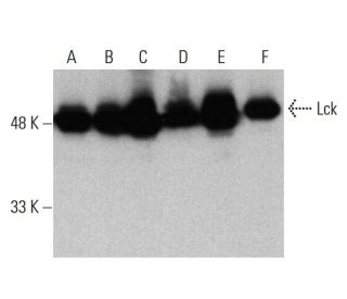
Lck Antibody (3A5): sc-433
- Lck Antibody (3A5) is a mouse monoclonal IgG2b κ Lck antibody, cited in 313 publications, provided at 200 µg/ml
- raised against amino acids 1-225 of Lck of mouse origin
- Lck Antibody (3A5) is recommended for detection of Lck p56 of mouse, rat and human origin by WB, IP, IF, IHC(P) and FCM
- Anti-Lck Antibody (3A5) is available conjugated to agarose for IP; HRP for WB, IHC(P) and ELISA; and to either phycoerythrin or FITC for IF, IHC(P) and FCM
- also available conjugated to Alexa Fluor® 488, Alexa Fluor® 546, Alexa Fluor® 594 or Alexa Fluor® 647 for WB (RGB), IF, IHC(P) and FCM, and for use with RGB fluorescent imaging systems, such as iBright™ FL1000, FluorChem™, Typhoon, Azure and other comparable systems
- also available conjugated to Alexa Fluor® 680 or Alexa Fluor® 790 for WB (NIR), IF and FCM; for use with Near-Infrared (NIR) detection systems, such as LI-COR®Odyssey®, iBright™ FL1000, FluorChem™, Typhoon, Azure and other comparable systems
- m-IgG2b BP-HRP and m-IgGκ BP-HRP are the preferred secondary detection reagents for Lck Antibody (3A5) for WB and IHC(P) applications. These reagents are now offered in bundles with Lck Antibody (3A5) (see ordering information below).

QUICK LINKS
SEE ALSO...
Lck Antibody (3A5) is a mouse monoclonal IgG2b kappa light chain antibody that detects Lck protein of mouse, rat, and human origin by western blotting (WB), immunoprecipitation (IP), immunofluorescence (IF), immunohistochemistry, and flow cytometry (FCM). Lck (3A5) antibody is available in both non-conjugated and various conjugated forms, including agarose, horseradish peroxidase (HRP), phycoerythrin (PE), fluorescein isothiocyanate (FITC), and multiple Alexa Fluor® conjugates. Lck protein, a member of the Src family of tyrosine kinases, plays a crucial role in T cell signaling, particularly in the activation of T cell antigen receptors (TCRs). This activation is vital for the immune response, as it triggers downstream signaling pathways that lead to T cell proliferation, differentiation, and cytokine production. Lck protein is primarily localized in the plasma membrane of T cells, where it associates with the TCR complex, ensuring that it is positioned to effectively transmit signals upon receptor engagement. The human Lck gene is located on chromosome 1p35.1 and encodes a 509 amino acid protein, which is essential for the regulation of immune responses. By understanding the function and localization of Lck, researchers can better appreciate its significance in T cell biology and its potential implications in immunological disorders.
Alexa Fluor® is a trademark of Molecular Probes Inc., OR., USA
LI-COR® and Odyssey® are registered trademarks of LI-COR Biosciences
Lck Antibody (3A5) References:
- Differential T-cell antigen receptor signaling mediated by the Src family kinases Lck and Fyn. | Denny, MF., et al. 2000. Mol Cell Biol. 20: 1426-35. PMID: 10648627
- Kinases of the Src family: structure and functions. | Tatosyan, AG. and Mizenina, OA. 2000. Biochemistry (Mosc). 65: 49-58. PMID: 10702640
- Src kinase-mediated signaling in leukocytes. | Korade-Mirnics, Z. and Corey, SJ. 2000. J Leukoc Biol. 68: 603-13. PMID: 11073097
- Selected glimpses into the activation and function of Src kinase. | Bjorge, JD., et al. 2000. Oncogene. 19: 5620-35. PMID: 11114743
- SRC family kinases mediate epithelial Na+ channel inhibition by endothelin. | Gilmore, ES., et al. 2001. J Biol Chem. 276: 42610-7. PMID: 11560932
- Asparagine enhances LCK signalling to potentiate CD8+ T-cell activation and anti-tumour responses. | Wu, J., et al. 2021. Nat Cell Biol. 23: 75-86. PMID: 33420490
- LAG3 associates with TCR-CD3 complexes and suppresses signaling by driving co-receptor-Lck dissociation. | Guy, C., et al. 2022. Nat Immunol. 23: 757-767. PMID: 35437325
- LCK inhibition downregulates YAP activity and is therapeutic in patient-derived models of cholangiocarcinoma. | Conboy, CB., et al. 2023. J Hepatol. 78: 142-152. PMID: 36162702
- CD28-CAR-T cell activation through FYN kinase signaling rather than LCK enhances therapeutic performance. | Wu, L., et al. 2023. Cell Rep Med. 4: 100917. PMID: 36696897
- A partial human LCK defect causes a T cell immunodeficiency with intestinal inflammation. | Lui, VG., et al. 2024. J Exp Med. 221: PMID: 37962568
- Genetic organization of human proto-oncogenes. | Sakaguchi, AY. 1983. Prog Clin Biol Res. 119: 93-103. PMID: 6191340
- Insights into Src kinase functions: structural comparisons. | Williams, JC., et al. 1998. Trends Biochem Sci. 23: 179-84. PMID: 9612082
Ordering Information
| Product Name | Catalog # | UNIT | Price | Qty | FAVORITES | |
Lck Antibody (3A5) | sc-433 | 200 µg/ml | $322.00 | |||
Lck Antibody (3A5): m-IgGκ BP-HRP Bundle | sc-520408 | 200 µg Ab, 40 µg BP | $361.00 | |||
Lck Antibody (3A5): m-IgG2b BP-HRP Bundle | sc-548825 | 200 µg Ab; 10 µg BP | $361.00 | |||
Lck Antibody (3A5) AC | sc-433 AC | 500 µg/ml, 25% agarose | $424.00 | |||
Lck Antibody (3A5) HRP | sc-433 HRP | 200 µg/ml | $322.00 | |||
Lck Antibody (3A5) FITC | sc-433 FITC | 200 µg/ml | $336.00 | |||
Lck Antibody (3A5) PE | sc-433 PE | 200 µg/ml | $349.00 | |||
Lck Antibody (3A5) Alexa Fluor® 488 | sc-433 AF488 | 200 µg/ml | $364.00 | |||
Lck Antibody (3A5) Alexa Fluor® 546 | sc-433 AF546 | 200 µg/ml | $364.00 | |||
Lck Antibody (3A5) Alexa Fluor® 594 | sc-433 AF594 | 200 µg/ml | $364.00 | |||
Lck Antibody (3A5) Alexa Fluor® 647 | sc-433 AF647 | 200 µg/ml | $364.00 | |||
Lck Antibody (3A5) Alexa Fluor® 680 | sc-433 AF680 | 200 µg/ml | $364.00 | |||
Lck Antibody (3A5) Alexa Fluor® 790 | sc-433 AF790 | 200 µg/ml | $364.00 |