
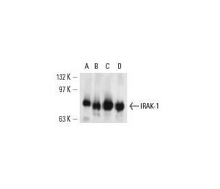
IRAK-1 Antibody (B-5): sc-55530
- IRAK-1 Antibody (B-5) is a mouse monoclonal IgG2b κ, cited in 7 publications, provided at 200 µg/ml
- raised against amino acids 440-712 mapping at the C-terminus of IRAK-1 of human origin
- recommended for detection of IRAK-1 of human origin by WB, IP, IF and ELISA
- See IRAK-1 (F-4): sc-5288 for IRAK-1 antibody conjugates, including AC, HRP, FITC, PE, Alexa Fluor® 488, 594, 647, 680 and 790.
- m-IgG Fc BP-HRP and m-IgGκ BP-HRP are the preferred secondary detection reagents for IRAK-1 Antibody (B-5) for WB applications. These reagents are now offered in bundles with IRAK-1 Antibody (B-5) (see ordering information below).

IRAK-1 Antibody (B-5) is a mouse monoclonal IgG2b antibody that detects IRAK-1 in human samples through applications such as western blotting (WB), immunoprecipitation (IP), immunofluorescence (IF), and enzyme-linked immunosorbent assay (ELISA). IRAK-1, or IL-1 receptor-associated kinase 1, plays a crucial role in the signaling pathways activated by interleukin-1 (IL-1), particularly in the activation of the NF-kB pathway, which is essential for regulating immune responses and inflammation. Located primarily in the cytoplasm, IRAK-1 is recruited to the IL-1 receptor complex upon ligand binding, where IRAK-1 undergoes phosphorylation and activation. This process is vital as IRAK-1 leads to the downstream activation of various transcription factors, including NF-kB, which in turn regulates the expression of numerous pro-inflammatory cytokines. The proper functioning of IRAK-1 is critical for maintaining immune homeostasis, and dysregulation of IRAK-1 activity has been implicated in various inflammatory diseases and conditions, making IRAK-1 a significant target for therapeutic intervention. Anti-IRAK-1 antibody (B-5) is an invaluable tool for researchers studying the intricate mechanisms of immune signaling and the pathophysiology of related diseases.
Alexa Fluor® is a trademark of Molecular Probes Inc., OR., USA
LI-COR® and Odyssey® are registered trademarks of LI-COR Biosciences
IRAK-1 Antibody (B-5) References:
- Translocation of the interleukin-1 receptor-associated kinase-1 (IRAK-1) into the nucleus. | Böl, G., et al. 2000. FEBS Lett. 477: 73-8. PMID: 10899313
- The death domain of IRAK-1: an oligomerization domain mediating interactions with MyD88, Tollip, IRAK-1, and IRAK-4. | Neumann, D., et al. 2007. Biochem Biophys Res Commun. 354: 1089-94. PMID: 17276401
- Leishmania-induced IRAK-1 inactivation is mediated by SHP-1 interacting with an evolutionarily conserved KTIM motif. | Abu-Dayyeh, I., et al. 2008. PLoS Negl Trop Dis. 2: e305. PMID: 19104650
- Regulation of IRAK-1 activation by its C-terminal domain. | Nguyen, T., et al. 2009. Cell Signal. 21: 719-26. PMID: 19166926
- Gender dependent importance of IRAK-1 in dextran sulfate sodium induced colitis. | Berglund, M., et al. 2009. Cell Immunol. 259: 27-32. PMID: 19540456
- IRAK-1 contributes to lipopolysaccharide-induced reactive oxygen species generation in macrophages by inducing NOX-1 transcription and Rac1 activation and suppressing the expression of antioxidative enzymes. | Maitra, U., et al. 2009. J Biol Chem. 284: 35403-11. PMID: 19850916
- Interleukin-1 Receptor-Associated Kinase-1 (IRAK-1) functionally associates with PKCepsilon and VASP in the regulation of macrophage migration. | Gan, L. and Li, L. 2010. Mol Immunol. 47: 1278-82. PMID: 20044140
- Differential regulation of interleukin-1 receptor-associated kinase-1 (IRAK-1) and IRAK-2 by microRNA-146a and NF-kappaB in stressed human astroglial cells and in Alzheimer disease. | Cui, JG., et al. 2010. J Biol Chem. 285: 38951-60. PMID: 20937840
- Increased expression of the interleukin-1 receptor-associated kinase (IRAK)-1 is associated with adipose tissue inflammatory state in obesity. | Ahmad, R., et al. 2015. Diabetol Metab Syndr. 7: 71. PMID: 26312071
- Inherited human IRAK-1 deficiency selectively impairs TLR signaling in fibroblasts. | Della Mina, E., et al. 2017. Proc Natl Acad Sci U S A. 114: E514-E523. PMID: 28069966
Ordering Information
| Product Name | Catalog # | UNIT | Price | Qty | FAVORITES | |
IRAK-1 Antibody (B-5) | sc-55530 | 200 µg/ml | $322.00 | |||
IRAK-1 Antibody (B-5): m-IgG Fc BP-HRP Bundle | sc-537064 | 200 µg Ab; 10 µg BP | $361.00 | |||
IRAK-1 Antibody (B-5): m-IgGκ BP-HRP Bundle | sc-534250 | 200 µg Ab; 40 µg BP | $361.00 |