")
HSP 40 Antibody (B-3): sc-398766
- HSP 40 Antibody (B-3) is a mouse monoclonal IgG1 κ HSP 40 antibody, cited in 8 publications, provided at 200 µg/ml
- raised against amino acids 241-340 mapping at the C-terminus of HSP 40 protein 1 of human origin
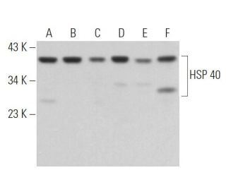
- HSP 40 Antibody (B-3) is recommended for detection of HSP 40 subfamily B member 1 of mouse, rat and human origin by WB, IP, IF and ELISA; also reactive with additional species, including and canine and bovine
- Anti-HSP 40 Antibody (B-3) is available conjugated to agarose for IP; HRP for WB, IHC(P) and ELISA; and to either phycoerythrin or FITC for IF, IHC(P) and FCM
- also available conjugated to Alexa Fluor® 488, Alexa Fluor® 546, Alexa Fluor® 594 or Alexa Fluor® 647 for WB (RGB), IF, IHC(P) and FCM, and for use with RGB fluorescent imaging systems, such as iBright™ FL1000, FluorChem™, Typhoon, Azure and other comparable systems
- also available conjugated to Alexa Fluor® 680 or Alexa Fluor® 790 for WB (NIR), IF and FCM; for use with Near-Infrared (NIR) detection systems, such as LI-COR®Odyssey®, iBright™ FL1000, FluorChem™, Typhoon, Azure and other comparable systems
- m-IgG Fc BP-HRP and m-IgG1 BP-HRP are the preferred secondary detection reagents for HSP 40 Antibody (B-3) for WB applications. These reagents are now offered in bundles with HSP 40 Antibody (B-3) (see ordering information below).

HSP 40 Antibody (B-3) is a mouse monoclonal IgG1 kappa light chain antibody that detects HSP 40 protein of mouse, rat, and human origin by western blotting (WB), immunoprecipitation (IP), immunofluorescence (IF), and enzyme-linked immunosorbent assay (ELISA). HSP 40 (B-3) antibody is available in both non-conjugated and various conjugated forms, including agarose, horseradish peroxidase (HRP), phycoerythrin (PE), fluorescein isothiocyanate (FITC), and multiple Alexa Fluor® conjugates. Heat shock protein 40 (HSP 40), also known as DnaJ, plays a crucial role in cellular stress responses by functioning as a molecular chaperone. HSP 40 interacts with HSP 70 through its J-domain, enhancing the ATPase activity of HSP 70, which is vital for protein folding and preventing aggregation of misfolded proteins. HSP 40 is predominantly located in the cytoplasm but translocates to the nucleus and nucleoli in response to heat shock, highlighting its importance in cellular adaptation to stress. This dynamic localization is essential for the regulation of protein homeostasis and the maintenance of cellular function under stress conditions. With five known family members, HSP 40 is integral to the assembly and disassembly of protein complexes, ensuring proper protein maturation and function.
Alexa Fluor® is a trademark of Molecular Probes Inc., OR., USA
LI-COR® and Odyssey® are registered trademarks of LI-COR Biosciences
HSP 40 Antibody (B-3) References:
- Heat shock protein (Hsp) 40 mutants inhibit Hsp70 in mammalian cells. | Michels, AA., et al. 1999. J Biol Chem. 274: 36757-63. PMID: 10593983
- Mammalian HSP40/DNAJ homologs: cloning of novel cDNAs and a proposal for their classification and nomenclature. | Ohtsuka, K. and Hata, M. 2000. Cell Stress Chaperones. 5: 98-112. PMID: 11147971
- Specificity of class II Hsp40 Sis1 in maintenance of yeast prion [RNQ+]. | Lopez, N., et al. 2003. Mol Biol Cell. 14: 1172-81. PMID: 12631732
- Expression of heat shock protein (Hsp) 70 and Hsp 40 in colorectal cancer. | Kanazawa, Y., et al. 2003. Med Oncol. 20: 157-64. PMID: 12835518
- Novel role of HSP40/DNAJ in the regulation of HIV-1 replication. | Urano, E., et al. 2013. J Acquir Immune Defic Syndr. 64: 154-62. PMID: 24047968
- The endoplasmic reticulum HSP40 co-chaperone ERdj3/DNAJB11 assembles and functions as a tetramer. | Chen, KC., et al. 2017. EMBO J. 36: 2296-2309. PMID: 28655754
- Targeting the Hsp40/Hsp70 Chaperone Axis as a Novel Strategy to Treat Castration-Resistant Prostate Cancer. | Moses, MA., et al. 2018. Cancer Res. 78: 4022-4035. PMID: 29764864
- Cotranslocation and colocalization of hsp40 (DnaJ) with hsp70 (DnaK) in mammalian cells. | Yamane, M., et al. 1995. Cell Struct Funct. 20: 157-66. PMID: 7641298
- Heat-shock protein 40, a novel predictor of thermotolerance in murine cells. | Kaneko, R., et al. 1995. Radiat Res. 142: 91-7. PMID: 7899564
- Effect of ATP on the release of hsp 70 and hsp 40 from the nucleus in heat-shocked HeLa cells. | Ohtsuka, K., et al. 1993. Exp Cell Res. 209: 357-66. PMID: 8262154
- Isolation of a new member of DnaJ-like heat shock protein 40 (Hsp40) from human liver. | Hoe, KL., et al. 1998. Biochim Biophys Acta. 1383: 4-8. PMID: 9546042
- Presence of molecular chaperones, heat shock cognate (Hsc) 70 and heat shock proteins (Hsp) 40, in the postsynaptic structures of rat brain. | Suzuki, T., et al. 1999. Brain Res. 816: 99-110. PMID: 9878698
Ordering Information
| Product Name | Catalog # | UNIT | Price | Qty | FAVORITES | |
HSP 40 Antibody (B-3) | sc-398766 | 200 µg/ml | $322.00 | |||
HSP 40 Antibody (B-3): m-IgG Fc BP-HRP Bundle | sc-527809 | 200 µg Ab; 10 µg BP | $361.00 | |||
HSP 40 Antibody (B-3): m-IgG1 BP-HRP Bundle | sc-533182 | 200 µg Ab; 20 µg BP | $361.00 | |||
HSP 40 Antibody (B-3) AC | sc-398766 AC | 500 µg/ml, 25% agarose | $424.00 | |||
HSP 40 Antibody (B-3) HRP | sc-398766 HRP | 200 µg/ml | $322.00 | |||
HSP 40 Antibody (B-3) FITC | sc-398766 FITC | 200 µg/ml | $336.00 | |||
HSP 40 Antibody (B-3) PE | sc-398766 PE | 200 µg/ml | $349.00 | |||
HSP 40 Antibody (B-3) Alexa Fluor® 488 | sc-398766 AF488 | 200 µg/ml | $364.00 | |||
HSP 40 Antibody (B-3) Alexa Fluor® 546 | sc-398766 AF546 | 200 µg/ml | $364.00 | |||
HSP 40 Antibody (B-3) Alexa Fluor® 594 | sc-398766 AF594 | 200 µg/ml | $364.00 | |||
HSP 40 Antibody (B-3) Alexa Fluor® 647 | sc-398766 AF647 | 200 µg/ml | $364.00 | |||
HSP 40 Antibody (B-3) Alexa Fluor® 680 | sc-398766 AF680 | 200 µg/ml | $364.00 | |||
HSP 40 Antibody (B-3) Alexa Fluor® 790 | sc-398766 AF790 | 200 µg/ml | $364.00 |