")
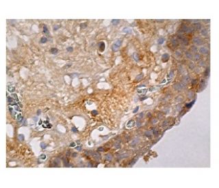
: sc-271918. Immunoperoxidase staining of formalin fixed, paraffin-embedded human urinary bladder tissue showing cytoplasmic staining of glandular cells.")
: sc-271918. Western blot analysis of human recombinant GnRH I fusion protein.")
GnRH I Antibody (A-4): sc-271918
- GnRH I Antibody (A-4) is a mouse monoclonal IgG2b κ GnRH I antibody, cited in 1 publications, provided at 200 µg/ml
- raised against amino acids 1-92 representing full length Progonadoliberin I of human origin
- GnRH I Antibody (A-4) is recommended for detection of Progonadoliberin I precursor and Gonadoliberin I and GnRH-associated peptide I active peptides of human origin by WB, IP, IF, IHC(P) and ELISA
- Anti-GnRH I Antibody (A-4) is available conjugated to agarose for IP; HRP for WB, IHC(P) and ELISA; and to either phycoerythrin or FITC for IF, IHC(P) and FCM
- also available conjugated to Alexa Fluor® 488, Alexa Fluor® 546, Alexa Fluor® 594 or Alexa Fluor® 647 for WB (RGB), IF, IHC(P) and FCM, and for use with RGB fluorescent imaging systems, such as iBright™ FL1000, FluorChem™, Typhoon, Azure and other comparable systems
- also available conjugated to Alexa Fluor® 680 or Alexa Fluor® 790 for WB (NIR), IF and FCM; for use with Near-Infrared (NIR) detection systems, such as LI-COR®Odyssey®, iBright™ FL1000, FluorChem™, Typhoon, Azure and other comparable systems
- m-IgG Fc BP-HRP, m-IgG2b BP-HRP and m-IgGκ BP-HRP are the preferred secondary detection reagents for GnRH I Antibody (A-4) for WB and IHC(P) applications. These reagents are now offered in bundles with GnRH I Antibody (A-4) (see ordering information below).

QUICK LINKS
GnRH I Antibody (A-4) is a mouse monoclonal IgG2b kappa light chain antibody that detects GnRH I protein of human origin by western blotting (WB), immunoprecipitation (IP), immunofluorescence (IF), immunohistochemistry with paraffin-embedded sections (IHCP), and enzyme-linked immunosorbent assay (ELISA). Anti-GnRH I antibody (A-4) is available in both non-conjugated and various conjugated forms, including agarose, horseradish peroxidase (HRP), phycoerythrin (PE), fluorescein isothiocyanate (FITC), and multiple Alexa Fluor® conjugates. GnRH I plays a crucial role in human reproduction through function in the hypothalamic-pituitary-gonadal axis, which is established early in fetal development. As a decapeptide, GnRH I is synthesized by hypothalamic neurons and released in a pulsatile manner into the capillary plexus of the median eminence, where GnRH I stimulates the secretion of luteinizing hormone and follicle-stimulating hormone from gonadotropic cells in the anterior pituitary. This regulation is vital for reproductive health, as GnRH I influences the development and function of the gonads. GnRH I is expressed in the acrosomal region of human sperm and in anterior pituitary tissue, as well as in certain cancer cells, indicating broader biological significance. A second form, GnRH II, primarily expressed outside the brain, suggests these peptides may have distinct and complementary roles in various physiological processes. The genes encoding GnRH-I and -II are located on human chromosomes 8p21-p11.2 and 20p13, respectively, highlighting their evolutionary importance.
Alexa Fluor® is a trademark of Molecular Probes Inc., OR., USA
LI-COR® and Odyssey® are registered trademarks of LI-COR Biosciences
Ordering Information
| Product Name | Catalog # | UNIT | Price | Qty | FAVORITES | |
GnRH I Antibody (A-4) | sc-271918 | 200 µg/ml | $322.00 | |||
GnRH I Antibody (A-4): m-IgG Fc BP-HRP Bundle | sc-529287 | 200 µg Ab; 10 µg BP | $361.00 | |||
GnRH I Antibody (A-4): m-IgGκ BP-HRP Bundle | sc-522101 | 200 µg Ab, 40 µg BP | $361.00 | |||
GnRH I Antibody (A-4): m-IgG2b BP-HRP Bundle | sc-550089 | 200 µg Ab; 10 µg BP | $361.00 | |||
GnRH I Antibody (A-4) AC | sc-271918 AC | 500 µg/ml, 25% agarose | $424.00 | |||
GnRH I Antibody (A-4) HRP | sc-271918 HRP | 200 µg/ml | $322.00 | |||
GnRH I Antibody (A-4) FITC | sc-271918 FITC | 200 µg/ml | $336.00 | |||
GnRH I Antibody (A-4) PE | sc-271918 PE | 200 µg/ml | $349.00 | |||
GnRH I Antibody (A-4) Alexa Fluor® 488 | sc-271918 AF488 | 200 µg/ml | $364.00 | |||
GnRH I Antibody (A-4) Alexa Fluor® 546 | sc-271918 AF546 | 200 µg/ml | $364.00 | |||
GnRH I Antibody (A-4) Alexa Fluor® 594 | sc-271918 AF594 | 200 µg/ml | $364.00 | |||
GnRH I Antibody (A-4) Alexa Fluor® 647 | sc-271918 AF647 | 200 µg/ml | $364.00 | |||
GnRH I Antibody (A-4) Alexa Fluor® 680 | sc-271918 AF680 | 200 µg/ml | $364.00 | |||
GnRH I Antibody (A-4) Alexa Fluor® 790 | sc-271918 AF790 | 200 µg/ml | $364.00 |