")
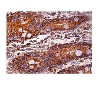
: sc-166056. Immunoperoxidase staining of formalin fixed, paraffin-embedded human duodenum tissue showing cytoplasmic staining of glandular cells.")
: sc-166056. Western blot analysis of GCAP2 expression in non-transfected: sc-117752 (A) and mouse GCAP2 transfected: sc-120436 (B) 293T whole cell lysates.")
GCAP2 Antibody (G-10): sc-166056
- GCAP2 Antibody (G-10) is a mouse monoclonal IgG2b κ GCAP2 antibody, cited in 2 publications, provided at 200 µg/ml
- raised against amino acids 130-201 mapping at the C-terminus of GCAP2 of mouse origin
- GCAP2 Antibody (G-10) is recommended for detection of GCAP2 of mouse, rat and human origin by WB, IP, IF, IHC(P) and ELISA
- Anti-GCAP2 Antibody (G-10) is available conjugated to agarose for IP; HRP for WB, IHC(P) and ELISA; and to either phycoerythrin or FITC for IF, IHC(P) and FCM
- also available conjugated to Alexa Fluor® 488, Alexa Fluor® 546, Alexa Fluor® 594 or Alexa Fluor® 647 for WB (RGB), IF, IHC(P) and FCM, and for use with RGB fluorescent imaging systems, such as iBright™ FL1000, FluorChem™, Typhoon, Azure and other comparable systems
- also available conjugated to Alexa Fluor® 680 or Alexa Fluor® 790 for WB (NIR), IF and FCM; for use with Near-Infrared (NIR) detection systems, such as LI-COR®Odyssey®, iBright™ FL1000, FluorChem™, Typhoon, Azure and other comparable systems
- m-IgG2b BP-HRP and m-IgGκ BP-HRP are the preferred secondary detection reagents for GCAP2 Antibody (G-10) for WB and IHC(P) applications. These reagents are now offered in bundles with GCAP2 Antibody (G-10) (see ordering information below).

QUICK LINKS
GCAP2 Antibody (G-10) is a mouse monoclonal IgG2b kappa light chain antibody that detects GCAP2 protein of mouse, rat, and human origin by western blotting (WB), immunoprecipitation (IP), immunofluorescence (IF), immunohistochemistry, and enzyme-linked immunosorbent assay (ELISA). GCAP2 (G-10) antibody is available in both non-conjugated and various conjugated forms, including agarose, horseradish peroxidase (HRP), phycoerythrin (PE), fluorescein isothiocyanate (FITC), and multiple Alexa Fluor® conjugates. GCAP2 plays a crucial role in the retina, specifically in the regulation of phototransduction, which is essential for converting light signals into visual information. GCAP2 is primarily located in the outer segments of rod photoreceptors, where GCAP2 interacts with guanylate cyclase to modulate cGMP levels in response to changes in intracellular calcium concentrations. This regulation is vital for the recovery of photoreceptors after exposure to light, ensuring that vision can adapt to varying light conditions. GCAP2 is a product of gene duplication and is closely related to GCAP1, with both proteins sharing over 90% similarity in their amino acid sequences and three conserved calcium-binding sites. GCAP1 and GCAP2 genes are located on human chromosome 6p21.1, highlighting their evolutionary significance and functional importance in visual signaling pathways.
Alexa Fluor® is a trademark of Molecular Probes Inc., OR., USA
LI-COR® and Odyssey® are registered trademarks of LI-COR Biosciences
GCAP2 Antibody (G-10) References:
- Conformational changes in guanylyl cyclase-activating protein 1 (GCAP1) and its tryptophan mutants as a function of calcium concentration. | Sokal, I., et al. 1999. J Biol Chem. 274: 19829-37. PMID: 10391927
- Guanylyl cyclase activating protein. A calcium-sensitive regulator of phototransduction. | Gorczyca, WA., et al. 1995. J Biol Chem. 270: 22029-36. PMID: 7665624
- Molecular characterization of human and mouse photoreceptor guanylate cyclase-activating protein (GCAP) and chromosomal localization of the human gene. | Subbaraya, I., et al. 1994. J Biol Chem. 269: 31080-9. PMID: 7983048
- Localization of guanylate cyclase-activating protein 2 in mammalian retinas. | Otto-Bruc, A., et al. 1997. Proc Natl Acad Sci U S A. 94: 4727-32. PMID: 9114059
- The human GCAP1 and GCAP2 genes are arranged in a tail-to-tail array on the short arm of chromosome 6 (p21.1). | Surguchov, A., et al. 1997. Genomics. 39: 312-22. PMID: 9119368
- Changes in biological activity and folding of guanylate cyclase-activating protein 1 as a function of calcium. | Rudnicka-Nawrot, M., et al. 1998. Biochemistry. 37: 248-57. PMID: 9425045
Ordering Information
| Product Name | Catalog # | UNIT | Price | Qty | FAVORITES | |
GCAP2 Antibody (G-10) | sc-166056 | 200 µg/ml | $322.00 | |||
GCAP2 Antibody (G-10): m-IgGκ BP-HRP Bundle | sc-521498 | 200 µg Ab, 40 µg BP | $361.00 | |||
GCAP2 Antibody (G-10): m-IgG2b BP-HRP Bundle | sc-548915 | 200 µg Ab; 10 µg BP | $361.00 | |||
GCAP2 Antibody (G-10) AC | sc-166056 AC | 500 µg/ml, 25% agarose | $424.00 | |||
GCAP2 Antibody (G-10) HRP | sc-166056 HRP | 200 µg/ml | $322.00 | |||
GCAP2 Antibody (G-10) FITC | sc-166056 FITC | 200 µg/ml | $336.00 | |||
GCAP2 Antibody (G-10) PE | sc-166056 PE | 200 µg/ml | $349.00 | |||
GCAP2 Antibody (G-10) Alexa Fluor® 488 | sc-166056 AF488 | 200 µg/ml | $364.00 | |||
GCAP2 Antibody (G-10) Alexa Fluor® 546 | sc-166056 AF546 | 200 µg/ml | $364.00 | |||
GCAP2 Antibody (G-10) Alexa Fluor® 594 | sc-166056 AF594 | 200 µg/ml | $364.00 | |||
GCAP2 Antibody (G-10) Alexa Fluor® 647 | sc-166056 AF647 | 200 µg/ml | $364.00 | |||
GCAP2 Antibody (G-10) Alexa Fluor® 680 | sc-166056 AF680 | 200 µg/ml | $364.00 | |||
GCAP2 Antibody (G-10) Alexa Fluor® 790 | sc-166056 AF790 | 200 µg/ml | $364.00 |