")
G3BP1抗体(H-10): sc-365338
- G3BP1抗体 H-10はマウスモノクローナルIgG1G3BP1 抗体 です。200 µg/mlで提供
- アミノ酸 158-251 マッピングに対して調製された human 由来のアポトーシスタンパク質 1 の内部領域内の G3BP1
- G3BP1抗体 (H-10) human由来のG3BP1 WB, IP, IF, IHC(P) と ELISAでの検出にはお勧めします
- 抗 G3BP1 抗体 (H-10) は、IP 用には アガロース、WB、IHC(P)、ELISA 用には HRP、IF、IHC(P)、FCM 用には フィコエリスリン または FITC にそれぞれ結合したものが利用可能
- WB (RGB)、IF、IHC(P)、FCM、iBright™ FL1000、FluorChem™、Typhoon、Azureと他の同等システムでRGB蛍光イメージングシステム用のAlexa Fluor® 488、Alexa Fluor® 546、Alexa Fluor® 594 または Alexa Fluor® 647、に共役での利用可能です。
- WB (NIR)、IF、FCMとLI-COR®/Odyssey®、iBright™ FL1000、FluorChem™、Typhoon、Azureと他の同等システムで近赤外(NIR)検出法用のAlexa Fluor® 680 または Alexa Fluor® 790、に共役での利用可能です。
- m-IgG Fc BP-HRP、 1 BP-HRP">m-IgG1 BP-HRPおよびm-IgGκ BP-HRPは、G3BP1 Antibody (H-10) WBおよびIHC(P)アプリケーション用。 の二次検出試薬として推奨されています。これらの試薬は現在、G3BP1 Antibody (H-10) とバンドルして提供されています(下記の注文情報を参照)。

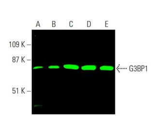
G3BP1 Antibody (H-10) は IgG1 κマウスモノクローナル G3BP1 抗体 (G3BP-1 抗体) で、ヒト由来の G3BP1 タンパク質を WB、IP、IF、IHC(P)、ELISA で検出します。G3BP1 Antibody (H-10) はノンコンジュゲート抗G3BP1抗体として、またアガロース、HRP、PE、FITC、Alexa Fluor® コンジュゲートなど複数のコンジュゲート抗G3BP1抗体としてご利用いただけます。G3BP1 (GTPase activating protein (SH3 domain) binding protein 1)は、G3BPまたはHDH-VIIIとしても知られ、ユビキタスに発現するタンパク質で、増殖細胞では細胞質に、非増殖細胞では核に局在します。いくつかのDNA巻き戻し酵素の一つであるG3BP1は、配列特異的なリン酸化依存性ヘリカーゼとして機能し、3′末端または5′末端がぶら下がった部分RNAおよびDNA二重鎖を巻き戻す。G3BP1はマグネシウムを補酵素として使い、ヘリカーゼ活性に加えて、3′-UTRのアデニン残基とシトシン残基の間でmRNAを切断するエンドリボヌクレアーゼとして働く。Rasシグナル伝達経路の一要素であるG3BP1は、増殖細胞においてRas GTPase-activating protein (Ras GAP)のSH3ドメインに結合し、それによって発生組織におけるRasシグナル伝達事象を制御している。DNA複製と細胞内のシグナル伝達経路の両方への関与により、G3BP1の発現は食道扁平上皮癌を含むいくつかの癌の病因に関与している。
Alexa Fluor® はMolecular Probes Inc., OR., USAの商標です。
LI-COR® and Odyssey® はLI-COR Biosciencesの登録商標です。
G3BP1抗体(H-10) 参考文献:
- Ras-GAP SH3ドメイン結合タンパク質(G3BP)は, 新規ヒトユビキチン特異的プロテアーゼであるUSP10のモジュレーターである。 | Soncini, C., et al. 2001. Oncogene. 20: 3869-79. PMID: 11439350
- RasGAP関連エンドリボヌクレアーゼG3Bp:選択的RNA分解とリン酸化依存性局在。 | Tourrière, H., et al. 2001. Mol Cell Biol. 21: 7747-60. PMID: 11604510
- RasGAP関連エンドリボヌクレアーゼG3BPはストレス顆粒を形成する。 | Tourrière, H., et al. 2003. J Cell Biol. 160: 823-31. PMID: 12642610
- ラスプーチン, かつてないほど乱暴に:G3BPのレビュー | Irvine, K., et al. 2004. Int J Dev Biol. 48: 1065-77. PMID: 15602692
- G3BP-1との相互作用, 真核生物翻訳開始因子2αのリン酸化誘導, 細胞質ストレス顆粒への進入, mRNAのサブセットとの選択的相互作用は, カプリン-1の特徴的な構造的特徴を媒介する。 | Solomon, S., et al. 2007. Mol Cell Biol. 27: 2324-42. PMID: 17210633
- Ras-GAP結合タンパク質(G3BP)との新規相互作用によるp53およびMDM2活性の調節。 | Kim, MM., et al. 2007. Oncogene. 26: 4209-15. PMID: 17297477
- 食道扁平上皮癌におけるG3BPとRhoCの発現と予後への影響。 | Zhang, HZ., et al. 2007. World J Gastroenterol. 13: 4126-30. PMID: 17696235
- 腫瘍促進におけるG3BP1の役割とメカニズム。 | Zhang, CH., et al. 2019. J Drug Target. 27: 300-305. PMID: 30207743
- 子宮内膜がん(EC)由来のG3BP1の過剰発現および変異体は、SPOP/ERα軸を介してECの腫瘍化および転移を促進する。 | Ge, Y., et al. 2023. Cell Commun Signal. 21: 303. PMID: 37904149
- DCAF7はUSP10をG3BP1の脱ユビキチン化の足場として働き、上咽頭癌の化学療法抵抗性と転移を促進する。 | Li, QJ., et al. 2024. Adv Sci (Weinh). 11: e2403262. PMID: 38973296
- Ras-GTPase活性化タンパク質SH3ドメイン結合タンパク質。 | Parker, F., et al. 1996. Mol Cell Biol. 16: 2561-9. PMID: 8649363
- ヒトDNAヘリカーゼVIII:G3BPタンパク質に対応するDNAおよびRNAヘリカーゼ。 | Costa, M., et al. 1999. Nucleic Acids Res. 27: 817-21. PMID: 9889278
注文情報
| 製品名 | カタログ # | 単位 | 価格 | 数量 | お気に入り | |
G3BP1 抗体 (H-10) | sc-365338 | 200 µg/ml | $322.00 | |||
G3BP1 (H-10): m-IgG Fc BP-HRP Bundle | sc-551727 | 200 µg Ab; 10 µg BP | $361.00 | |||
G3BP1 (H-10): m-IgGκ BP-HRP Bundle | sc-522213 | 200 µg Ab, 40 µg BP | $361.00 | |||
G3BP1 (H-10): m-IgG1 BP-HRP Bundle | sc-551728 | 200 µg Ab; 20 µg BP | $361.00 | |||
G3BP1 抗体 (H-10) AC | sc-365338 AC | 500 µg/ml, 25% agarose | $424.00 | |||
G3BP1 抗体 (H-10) HRP | sc-365338 HRP | 200 µg/ml | $322.00 | |||
G3BP1 抗体 (H-10) FITC | sc-365338 FITC | 200 µg/ml | $336.00 | |||
G3BP1 抗体 (H-10) PE | sc-365338 PE | 200 µg/ml | $349.00 | |||
G3BP1 抗体 (H-10) Alexa Fluor® 488 | sc-365338 AF488 | 200 µg/ml | $364.00 | |||
G3BP1 抗体 (H-10) Alexa Fluor® 546 | sc-365338 AF546 | 200 µg/ml | $364.00 | |||
G3BP1 抗体 (H-10) Alexa Fluor® 594 | sc-365338 AF594 | 200 µg/ml | $364.00 | |||
G3BP1 抗体 (H-10) Alexa Fluor® 647 | sc-365338 AF647 | 200 µg/ml | $364.00 | |||
G3BP1 抗体 (H-10) Alexa Fluor® 680 | sc-365338 AF680 | 200 µg/ml | $364.00 | |||
G3BP1 抗体 (H-10) Alexa Fluor® 790 | sc-365338 AF790 | 200 µg/ml | $364.00 |