")
EF-1 α1/2 Antibody (G-8): sc-377439
- EF-1 α1/2 Antibody (G-8) is a mouse monoclonal IgG1 κ EF-1 α1/2 antibody, cited in 3 publications, provided at 200 µg/ml
- raised against amino acids 163-462 mapping at the C-terminus of EF-1 α1 of human origin
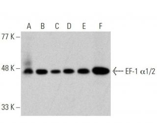
- EF-1 alpha 1/2 Antibody (G-8) is recommended for detection of EF-1 α1 and EF-1 α2 of mouse, rat, human and origin by WB, IP, IF, IHC(P) and ELISA; also reactive with additional species, including and equine, canine, bovine, porcine and avian
- Anti-EF-1 alpha 1/2 Antibody (G-8) is available conjugated to agarose for IP; HRP for WB, IHC(P) and ELISA; and to either phycoerythrin or FITC for IF, IHC(P) and FCM
- also available conjugated to Alexa Fluor® 488, Alexa Fluor® 546, Alexa Fluor® 594 or Alexa Fluor® 647 for WB (RGB), IF, IHC(P) and FCM, and for use with RGB fluorescent imaging systems, such as iBright™ FL1000, FluorChem™, Typhoon, Azure and other comparable systems
- also available conjugated to Alexa Fluor® 680 or Alexa Fluor® 790 for WB (NIR), IF and FCM; for use with Near-Infrared (NIR) detection systems, such as LI-COR®Odyssey®, iBright™ FL1000, FluorChem™, Typhoon, Azure and other comparable systems
- m-IgG Fc BP-HRP, m-IgG1 BP-HRP and m-IgGκ BP-HRP are the preferred secondary detection reagents for EF-1 α1/2 Antibody (G-8) for WB and IHC(P) applications. These reagents are now offered in bundles with EF-1 α1/2 Antibody (G-8) (see ordering information below).
EF-1 α1/2 Antibody (G-8) is a mouse monoclonal IgG1 kappa light chain antibody that detects EF-1 alpha 1/2 of mouse, rat, and human origin by western blotting (WB), immunoprecipitation (IP), immunofluorescence (IF), immunohistochemistry with paraffin-embedded sections (IHCP), and enzyme-linked immunosorbent assay (ELISA). Anti-EF-1 α1/2 antibody (G-8) is available in both non-conjugated and various conjugated forms, including agarose, horseradish peroxidase (HRP), phycoerythrin (PE), fluorescein isothiocyanate (FITC), and multiple Alexa Fluor® conjugates. The elongation factor-1 complex, consisting of EF-1 α1 and EF-1 α2, plays a crucial role in protein synthesis by facilitating the delivery of aminoacyl tRNAs to the ribosome, a process essential for translating genetic information into functional proteins. Proper functioning of EF-1 alpha ensures accurate and efficient protein synthesis, particularly important in rapidly dividing cells. EF-1 α1 is predominantly expressed in the brain, placenta, lung, liver, kidney, and pancreas, while EF-1 α2 shows high expression levels in the heart, brain, and skeletal muscle. Both isoforms localize to the nucleus and are part of the GTP-binding elongation factor family. The gene encoding EF-1 α2, located on human chromosome 20q13.3, has been implicated in ovarian cancer development, whereas the EF-1 α1 gene, found on chromosome 6Q14.1, is often recognized as an autoantigen in patients with Felty syndrome, a condition characterized by rheumatoid arthritis, splenomegaly, leukopenia, and increased infection risk.
Alexa Fluor® is a trademark of Molecular Probes Inc., OR., USA
LI-COR® and Odyssey® are registered trademarks of LI-COR Biosciences
EF-1 α1/2 Antibody (G-8) References:
- The role of protein elongation factor eEF1A2 in ovarian cancer. | Lee, JM. 2003. Reprod Biol Endocrinol. 1: 69. PMID: 14588074
- Retropseudogenes constitute the major part of the human elongation factor 1 alpha gene family. | Madsen, HO., et al. 1990. Nucleic Acids Res. 18: 1513-6. PMID: 2183196
- Dissecting the expression of EEF1A1/2 genes in human prostate cancer cells: the potential of EEF1A2 as a hallmark for prostate transformation and progression. | Scaggiante, B., et al. 2012. Br J Cancer. 106: 166-73. PMID: 22095224
- Eukaryotic translation elongation factor 1A induces anoikis by triggering cell detachment. | Itagaki, K., et al. 2012. J Biol Chem. 287: 16037-46. PMID: 22399298
- Inhibition of ER stress-mediated apoptosis in macrophages by nuclear-cytoplasmic relocalization of eEF1A by the HIV-1 Nef protein. | Abbas, W., et al. 2012. Cell Death Dis. 3: e292. PMID: 22476100
- Hepatitis B virus X protein blocks filamentous actin bundles by interaction with eukaryotic translation elongat ion factor 1 alpha 1. | Lin, WS., et al. 2012. J Med Virol. 84: 871-7. PMID: 22499008
- Molecular markers associated with nonepithelial ovarian cancer in formalin-fixed, paraffin-embedded specimens by genome wide expression profiling. | Vui-Kee, K., et al. 2012. Kaohsiung J Med Sci. 28: 243-50. PMID: 22531302
- Isolation and characterization of the human chromosomal gene for polypeptide chain elongation factor-1 alpha. | Uetsuki, T., et al. 1989. J Biol Chem. 264: 5791-8. PMID: 2564392
- The primary structure of the alpha subunit of human elongation factor 1. Structural aspects of guanine-nucleotide-binding sites. | Brands, JH., et al. 1986. Eur J Biochem. 155: 167-71. PMID: 3512269
- A novel 3'tRNA-derived fragment tRF-Val promotes proliferation and inhibits apoptosis by targeting EEF1A1 in gastric cancer. | Cui, H., et al. 2022. Cell Death Dis. 13: 471. PMID: 35585048
- Nanoparticles (NPs)-mediated lncBCMA silencing to promote eEF1A1 ubiquitination and suppress breast cancer growth and metastasis. | Yang, K., et al. 2023. Acta Pharm Sin B. 13: 3489-3502. PMID: 37655325
- KAT8-catalyzed lactylation promotes eEF1A2-mediated protein synthesis and colorectal carcinogenesis. | Xie, B., et al. 2024. Proc Natl Acad Sci U S A. 121: e2314128121. PMID: 38359291
Ordering Information
| Product Name | Catalog # | UNIT | Price | Qty | FAVORITES | |
EF-1 α1/2 Antibody (G-8) | sc-377439 | 200 µg/ml | $316.00 | |||
EF-1 α1/2 Antibody (G-8): m-IgG Fc BP-HRP Bundle | sc-530027 | 200 µg Ab; 10 µg BP | $354.00 | |||
EF-1 α1/2 Antibody (G-8): m-IgGκ BP-HRP Bundle | sc-523329 | 200 µg Ab, 40 µg BP | $354.00 | |||
EF-1 α1/2 Antibody (G-8): m-IgG1 BP-HRP Bundle | sc-543826 | 200 µg Ab; 20 µg BP | $354.00 | |||
EF-1 α1/2 Antibody (G-8) AC | sc-377439 AC | 500 µg/ml, 25% agarose | $416.00 | |||
EF-1 α1/2 Antibody (G-8) HRP | sc-377439 HRP | 200 µg/ml | $316.00 | |||
EF-1 α1/2 Antibody (G-8) FITC | sc-377439 FITC | 200 µg/ml | $330.00 | |||
EF-1 α1/2 Antibody (G-8) PE | sc-377439 PE | 200 µg/ml | $343.00 | |||
EF-1 α1/2 Antibody (G-8) Alexa Fluor® 488 | sc-377439 AF488 | 200 µg/ml | $357.00 | |||
EF-1 α1/2 Antibody (G-8) Alexa Fluor® 546 | sc-377439 AF546 | 200 µg/ml | $357.00 | |||
EF-1 α1/2 Antibody (G-8) Alexa Fluor® 594 | sc-377439 AF594 | 200 µg/ml | $357.00 | |||
EF-1 α1/2 Antibody (G-8) Alexa Fluor® 647 | sc-377439 AF647 | 200 µg/ml | $357.00 | |||
EF-1 α1/2 Antibody (G-8) Alexa Fluor® 680 | sc-377439 AF680 | 200 µg/ml | $357.00 | |||
EF-1 α1/2 Antibody (G-8) Alexa Fluor® 790 | sc-377439 AF790 | 200 µg/ml | $357.00 |