")
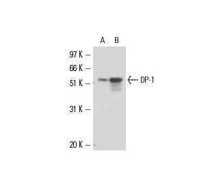
DP-1 Antibody (TFD-11): sc-56657
- DP-1 Antibody (TFD-11) is a mouse monoclonal IgG2b κ, cited in 2 publications, provided at 200 µg/ml
- raised against full length DP-1 of human origin
- recommended for detection of DP-1 of mouse, rat and human origin by WB, IP, IF, IHC(P) and ELISA
- m-IgG Fc BP-HRP and m-IgG2b BP-HRP are the preferred secondary detection reagents for DP-1 Antibody (TFD-11) for WB and IHC(P) applications. These reagents are now offered in bundles with DP-1 Antibody (TFD-11) (see ordering information below).

DP-1 Antibody (TFD-11) is a mouse monoclonal IgG2b antibody that detects DP-1 in mouse, rat, and human samples through various applications including western blotting (WB), immunoprecipitation (IP), immunofluorescence (IF), immunohistochemistry with paraffin-embedded sections (IHCP), and enzyme-linked immunosorbent assay (ELISA). DP-1, a crucial component of the E2F transcriptional regulatory complex, plays a significant role in cell cycle regulation by forming heterodimers with E2F-1, thereby facilitating the transcriptional activation of genes essential for the G1 and S phases of the cell cycle. The interaction between DP-1 and E2F-1 ensures the timely expression of genes that drive cell proliferation and division. Dysregulation of this complex can lead to uncontrolled cell growth, contributing to tumorigenesis. DP-1′s ability to bind with E2F-1 and other E2F family members, such as E2F-2 and E2F-3, underscores its importance in maintaining cellular homeostasis and highlights its potential as a target for therapeutic interventions in cancer and other proliferative disorders.
Alexa Fluor® is a trademark of Molecular Probes Inc., OR., USA
LI-COR® and Odyssey® are registered trademarks of LI-COR Biosciences
DP-1 Antibody (TFD-11) References:
- The DP-1 transcription factor is required for keratinocyte growth and epidermal stratification. | Chang, WY., et al. 2004. J Biol Chem. 279: 51343-53. PMID: 15448153
- Identification of significant regions of transcription factor DP-1 (TFDP-1) involved in stability/instability of the protein. | Arakawa, T., et al. 2010. Biochem Biophys Res Commun. 397: 345-9. PMID: 20513349
- U2 small nuclear RNA auxiliary factor 2, transcriptionally activated by the transcription factor Dp-1/E2F transcription factor 1 complex, enhances the growth and aerobic glycolysis of leiomyosarcoma cells. | Li, Y., et al. 2022. Bioengineered. 13: 10200-10212. PMID: 35502531
- Dispersion Function of a Protein, DP-1, Identified in Collimonas sp. D-25, for the Synthesis of Gold Nanoparticles. | Tang, D., et al. 2023. Chembiochem. 24: e202300221. PMID: 37232370
- Transcription factor Dp-1 knockdown downregulates thymidine kinase 1 expression to protect against proliferation and epithelial-mesenchymal transition in cervical cancer. | Wu, M. and Ye, M. 2023. Funct Integr Genomics. 23: 301. PMID: 37715794
- Functional synergy between DP-1 and E2F-1 in the cell cycle-regulating transcription factor DRTF1/E2F. | Bandara, LR., et al. 1993. EMBO J. 12: 4317-24. PMID: 8223441
- Heterodimerization of the transcription factors E2F-1 and DP-1 leads to cooperative trans-activation. | Helin, K., et al. 1993. Genes Dev. 7: 1850-61. PMID: 8405995
- E2F-1:DP-1 induces p53 and overrides survival factors to trigger apoptosis. | Hiebert, SW., et al. 1995. Mol Cell Biol. 15: 6864-74. PMID: 8524253
- Functional interaction between DP-1 and p53. | Sørensen, TS., et al. 1996. Mol Cell Biol. 16: 5888-95. PMID: 8816502
- Inhibition of E2F-4/DP-1-stimulated transcription by p202. | Choubey, D. and Gutterman, JU. 1997. Oncogene. 15: 291-301. PMID: 9233764
Ordering Information
| Product Name | Catalog # | UNIT | Price | Qty | FAVORITES | |
DP-1 Antibody (TFD-11) | sc-56657 | 200 µg/ml | $322.00 | |||
DP-1 Antibody (TFD-11): m-IgG Fc BP-HRP Bundle | sc-539035 | 200 µg Ab; 10 µg BP | $361.00 | |||
DP-1 Antibody (TFD-11): m-IgG2b BP-HRP Bundle | sc-549727 | 200 µg Ab; 10 µg BP | $361.00 |