")
CLN3 Antibody (C-1): sc-398192
- CLN3 Antibody (C-1) is a mouse monoclonal IgG2b κ CLN3 antibody, cited in 1 publications, provided at 200 µg/ml
- specific for an epitope mapping between amino acids 13-40 at the N-terminus of CLN3 of human origin
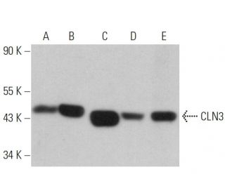
- CLN3 Antibody (C-1) is recommended for detection of CLN3 of mouse, rat and human origin by WB, IP, IF and ELISA
- Anti-CLN3 Antibody (C-1) is available conjugated to agarose for IP; HRP for WB, IHC(P) and ELISA; and to either phycoerythrin or FITC for IF, IHC(P) and FCM
- also available conjugated to Alexa Fluor® 488, Alexa Fluor® 546, Alexa Fluor® 594 or Alexa Fluor® 647 for WB (RGB), IF, IHC(P) and FCM, and for use with RGB fluorescent imaging systems, such as iBright™ FL1000, FluorChem™, Typhoon, Azure and other comparable systems
- also available conjugated to Alexa Fluor® 680 or Alexa Fluor® 790 for WB (NIR), IF and FCM; for use with Near-Infrared (NIR) detection systems, such as LI-COR®Odyssey®, iBright™ FL1000, FluorChem™, Typhoon, Azure and other comparable systems
- m-IgG Fc BP-HRP, m-IgG2b BP-HRP and m-IgGκ BP-HRP are the preferred secondary detection reagents for CLN3 Antibody (C-1) for WB applications. These reagents are now offered in bundles with CLN3 Antibody (C-1) (see ordering information below).

QUICK LINKS
CLN3 Antibody (C-1) is a mouse monoclonal IgG2b kappa light chain antibody that detects CLN3 protein of mouse, rat, and human origin by western blotting (WB), immunoprecipitation (IP), immunofluorescence (IF), and enzyme-linked immunosorbent assay (ELISA). CLN3 (C-1) antibody is available in both non-conjugated and various conjugated forms, including agarose, horseradish peroxidase (HRP), phycoerythrin (PE), fluorescein isothiocyanate (FITC), and multiple Alexa Fluor® conjugates. CLN3 protein is a highly glycosylated, hydrophobic protein consisting of 438 amino acids and features six transmembrane domains, which are crucial for function in the lysosomal membrane. This localization is significant as CLN3 plays a vital role in lysosomal function, potentially acting as a chaperone that assists in the proper folding and unfolding of other proteins, including subunit C of the ATP synthase complex. Mutations in CLN3 gene are linked to Batten disease, a recessively inherited neurodegenerative disorder characterized by lysosomal accumulation of hydrophobic materials, primarily affecting the ATP synthase subunit C. Batten disease represents the most prevalent form of a group of disorders known as neuronal ceroid lipofuscinoses (NCLs), with symptoms that include progressive vision loss, seizures, and psychomotor disturbances, underscoring the importance of CLN3 protein in maintaining cellular health and function.
Alexa Fluor® is a trademark of Molecular Probes Inc., OR., USA
LI-COR® and Odyssey® are registered trademarks of LI-COR Biosciences
CLN3 Antibody (C-1) References:
- Membrane trafficking and mitochondrial abnormalities precede subunit c deposition in a cerebellar cell model of juvenile neuronal ceroid lipofuscinosis. | Fossale, E., et al. 2004. BMC Neurosci. 5: 57. PMID: 15588329
- CLN3, the protein associated with batten disease: structure, function and localization. | Phillips, SN., et al. 2005. J Neurosci Res. 79: 573-83. PMID: 15657902
- Correlations between genotype, ultrastructural morphology and clinical phenotype in the neuronal ceroid lipofuscinoses. | Mole, SE., et al. 2005. Neurogenetics. 6: 107-26. PMID: 15965709
- Gene symbol: CLN3. Disease: Juvenile neuronal ceroid lipofuscinosis (Batten disease). | Leman, AR., et al. 2005. Hum Genet. 116: 544. PMID: 15991331
- Thalamocortical neuron loss and localized astrocytosis in the Cln3Deltaex7/8 knock-in mouse model of Batten disease. | Pontikis, CC., et al. 2005. Neurobiol Dis. 20: 823-36. PMID: 16006136
- Novel CLN3 mutation predicted to cause complete loss of protein function does not modify the classical JNCL phenotype. | Kwon, JM., et al. 2005. Neurosci Lett. 387: 111-4. PMID: 16087292
- Cell death pathways in juvenile Batten disease. | Persaud-Sawin, DA. and Boustany, RM. 2005. Apoptosis. 10: 973-85. PMID: 16151633
Ordering Information
| Product Name | Catalog # | UNIT | Price | Qty | FAVORITES | |
CLN3 Antibody (C-1) | sc-398192 | 200 µg/ml | $322.00 | |||
CLN3 Antibody (C-1): m-IgG Fc BP-HRP Bundle | sc-530716 | 200 µg Ab; 10 µg BP | $361.00 | |||
CLN3 Antibody (C-1): m-IgGκ BP-HRP Bundle | sc-524246 | 200 µg Ab, 40 µg BP | $361.00 | |||
CLN3 Antibody (C-1): m-IgG2b BP-HRP Bundle | sc-550217 | 200 µg Ab; 10 µg BP | $361.00 | |||
CLN3 Antibody (C-1) AC | sc-398192 AC | 500 µg/ml, 25% agarose | $424.00 | |||
CLN3 Antibody (C-1) HRP | sc-398192 HRP | 200 µg/ml | $322.00 | |||
CLN3 Antibody (C-1) FITC | sc-398192 FITC | 200 µg/ml | $336.00 | |||
CLN3 Antibody (C-1) PE | sc-398192 PE | 200 µg/ml | $349.00 | |||
CLN3 Antibody (C-1) Alexa Fluor® 488 | sc-398192 AF488 | 200 µg/ml | $364.00 | |||
CLN3 Antibody (C-1) Alexa Fluor® 546 | sc-398192 AF546 | 200 µg/ml | $364.00 | |||
CLN3 Antibody (C-1) Alexa Fluor® 594 | sc-398192 AF594 | 200 µg/ml | $364.00 | |||
CLN3 Antibody (C-1) Alexa Fluor® 647 | sc-398192 AF647 | 200 µg/ml | $364.00 | |||
CLN3 Antibody (C-1) Alexa Fluor® 680 | sc-398192 AF680 | 200 µg/ml | $364.00 | |||
CLN3 Antibody (C-1) Alexa Fluor® 790 | sc-398192 AF790 | 200 µg/ml | $364.00 | |||
CLN3 (C-1) Neutralizing Peptide | sc-398192 P | 100 µg/0.5 ml | $69.00 |