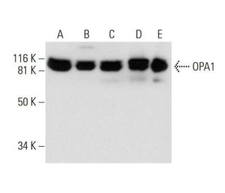
Date published: 2025-9-7

00800 4573 8000

SCBT Portrait Logo
Seach Input

OPA1 Anticorpo (D-9): sc-393296

4.0(22)
Escrever uma avaliaçãoFazer uma pergunta

Fichas de dados
  • OPA1 Anticorpo D-9é um anticorpo monoclonal produzido em camundongo IgG1 κ OPA1 anticorpo, citado em 62 publicações, fornecido em 200 µg/ml
  • Produzido contra aminoácidos 647-780 mapeamento dentro de uma região interna deOPA1 de human origem
  • OPA1 Anticorpo (D-9) é recomendado para a detecção de OPA1 de mouse, rat e human origem em WB, IP, IF e ELISA; Reage também com outras espécies, incluindo and equine, canine, bovine and porcine
  • Anticorpo anti-OPA1 O anticorpo anti-OPA1 (D-9) está disponível conjugado com agarose para IP; HRP para WB, IHC(P) e ELISA; e com fioeritrina ou FITC para IF, IHC(P) e FCM
  • também disponível conjugado com Alexa Fluor® 488, Alexa Fluor® 546, Alexa Fluor® 594 ou Alexa Fluor® 647 para WB (RGB), IF, IHC(P) e FCM, e para utilização com sistemas de imagiologia fluorescente RGB, tais como iBright™ FL1000, FluorChem™, Typhoon, Azure e outros sistemas comparáveis
  • também disponível conjugado com Alexa Fluor® 680 ou Alexa Fluor® 790 para WB (NIR), IF e FCM; para utilização com sistemas de deteção de infravermelhos próximos (NIR), tais como LI-COR®Odyssey®, iBright™ FL1000, FluorChem™, Typhoon, Azure e outros sistemas comparáveis
  • Entre em contato conosco e receba GRATUITAMENTE um amostra de 10 µg de OPA1 (D-9): sc-393296.
  • O m-IgG Fc BP-HRP e o m-IgGκ BP-HRP são os reagentes de deteção secundários preferidos para o anticorpo OPA1 (D-9) para aplicações WB. Estes reagentes são agora disponibilizados em conjuntos com o anticorpo OPA1 (D-9)(ver informações de encomenda abaixo).

LINKS RÁPIDOS

VEJA TAMBÉM

O anticorpo OPA1 (D-9) é um anticorpo monoclonal de camundongo IgG1 κ para OPA1 (também designado anticorpo para proteína mitocondrial semelhante à dinamina de 120 KDa, anticorpo para guanosina trifosfatase semelhante à dinamina, anticorpo para atrofia óptica 1 (autossômica dominante), Anticorpo de GTPase semelhante à dinamina mitocondrial, anticorpo de Proteína de Atrofia Óptica 1, anticorpo MGM1, anticorpo NPG, anticorpo NTG ou anticorpo BERHS) que detecta a proteína OPA1 de origem camundongo, rato e humana por WB, IP, IF e ELISA. O anticorpo OPA1 (D-9) está disponível na forma de anticorpo anti-OPA1 não conjugado, bem como em várias formas conjugadas de anticorpo anti-OPA1, incluindo agarose, HRP, PE, FITC e vários conjugados Alexa Fluor®. OPA1 (proteína do gene da atrofia óptica 1) pertence à família genética Dynamin. O gene que codifica a OPA1 localiza-se em 3q29, tem como alvo a mitocôndria e está envolvido na biogênese mitocondrial. Defeitos no OPA1 são uma das causas da atrofia óptica tipo 1. O OPA1 é expresso principalmente na retina, mas também pode ser expresso no cérebro, testículo, coração e músculos esqueléticos.

Para uso em exclusivo em pesquisa. Não se destina a uso em diagnostico e tratamento.

Alexa Fluor® é uma marca comercial da Molecular Probes Inc., OR., EUA

LI-COR® e Odyssey® são marcas registadas da LI-COR Biosciences

Referencias do OPA1 Anticorpo (D-9):

  1. O gene nuclear OPA1, que codifica uma proteína mitocondrial relacionada com a dinamina, está mutado na atrofia ótica dominante.  |  Delettre, C., et al. 2000. Nat Genet. 26: 207-10. PMID: 11017079
  2. Espectro, frequência e penetrância das mutações OPA1 na atrofia ótica dominante.  |  Toomes, C., et al. 2001. Hum Mol Genet. 10: 1369-78. PMID: 11440989
  3. Espectro de mutações e variantes de splicing no gene OPA1.  |  Delettre, C., et al. 2001. Hum Genet. 109: 584-91. PMID: 11810270
  4. Sublocalização diferencial das isoformas da proteína relacionada com a dinamina OPA1 nas mitocôndrias.  |  Satoh, M., et al. 2003. Biochem Biophys Res Commun. 300: 482-93. PMID: 12504110
  5. Papéis dos mediadores de fissão e fusão mitocondrial de mamíferos Fis1, Drp1 e Opa1 na apoptose.  |  Lee, YJ., et al. 2004. Mol Biol Cell. 15: 5001-11. PMID: 15356267
  6. OPA1 requer mitofusina 1 para promover a fusão mitocondrial.  |  Cipolat, S., et al. 2004. Proc Natl Acad Sci U S A. 101: 15927-32. PMID: 15509649
  7. OPA1, associado à atrofia ótica autossómica dominante, é amplamente expresso no cérebro humano.  |  Bette, S., et al. 2005. Acta Neuropathol. 109: 393-9. PMID: 15700187
  8. Refinamento do locus dominante da atrofia ótica (OPA1) para um intervalo de 1,4 cm no cromossoma 3q28-3q29, dentro de um contig YAC de 3 Mb.  |  Jonasdottir, A., et al. 1997. Hum Genet. 99: 115-20. PMID: 9003507

Informacoes sobre ordens

Nome do ProdutoNumero de CatalogoUNIDPrecoQdeFAVORITOS

OPA1 Anticorpo (D-9)

sc-393296
200 µg/ml
$316.00

Pacote do OPA1 (D-9): m-IgG Fc BP-HRP

sc-530422
200 µg Ab; 10 µg BP
$354.00

Pacote do OPA1 (D-9): m-IgGκ BP-HRP

sc-523893
200 µg Ab, 40 µg BP
$354.00

OPA1 Anticorpo (D-9) AC

sc-393296 AC
500 µg/ml, 25% agarose
$416.00

OPA1 Anticorpo (D-9) HRP

sc-393296 HRP
200 µg/ml
$316.00

OPA1 Anticorpo (D-9) FITC

sc-393296 FITC
200 µg/ml
$330.00

OPA1 Anticorpo (D-9) PE

sc-393296 PE
200 µg/ml
$343.00

OPA1 Anticorpo (D-9) Alexa Fluor® 488

sc-393296 AF488
200 µg/ml
$357.00

OPA1 Anticorpo (D-9) Alexa Fluor® 546

sc-393296 AF546
200 µg/ml
$357.00

OPA1 Anticorpo (D-9) Alexa Fluor® 594

sc-393296 AF594
200 µg/ml
$357.00

OPA1 Anticorpo (D-9) Alexa Fluor® 647

sc-393296 AF647
200 µg/ml
$357.00

OPA1 Anticorpo (D-9) Alexa Fluor® 680

sc-393296 AF680
200 µg/ml
$357.00

OPA1 Anticorpo (D-9) Alexa Fluor® 790

sc-393296 AF790
200 µg/ml
$357.00

I am currently looking for antibodies for OPA1 for immunofluorescence. I would like to ask if it is possible to get a trial sample to try? We have OPA1 ko cells so if it works, I could upload some photos and protocols to share. Many thanks in advance!

Asked by: Xiaotong
Please contact our Technical Service Department for more information on how to receive a free 10 ug sample of this monoclonal antibody.
Answered by: Technical Support
Date published: 2020-07-24

As I am unhappy with the current OPA1-antibody we are using (other manufacturer), I am looking to buy a new one. Would it be possible to get a free sample of this antibody for testing?

Asked by: Christiaan
Thank you for your question. Please contact Technical Service for information on how to receive a free 10 ug sample of this monoclonal antibody.
Answered by: Tech Service
Date published: 2020-02-06

8% separation gel, first antibody 1:100, 4℃,overnight. second antibody 1:1000,room temperature, 1 hour. But I think the band (130-250kDa) is not right.

Asked by: Anonymous
Thank you for your question. Please contact your local distributor or Technical Service directly for assistance regarding your WB results, we are happy to discuss your results with you.
Answered by: Tech Service
Date published: 2019-04-26

what buffer does the opa1 antibody work best in? I tried probing a blot of mouse heart tissue with opa1 at 1:1000 in 5%milk/ TBST and the blot is there is a lot of background. I checked the publications cited and they lack the buffer details

Asked by: Dr C
Thank you for your question. The optimal blocking buffer and antibody diluent depends on which type of Western blot detection method you are using. If you are using an HRP conjugated secondary for chemiluminescent detection, then we recommend Blotto, or 5% nonfat milk in TBST (1X TBST + 0.05% Tween-20) for blocking, and for incubating both primary and secondary antibodies. If you are using a fluorescent or NIR (Near-Infrared) detection method, for optimal results, use low fluorescence PVDF membranes, such as Immobilon®-FL PVDF: sc-516541, and UltraCruz® Blocking Reagent: sc-516214 for all blocking, primary antibody and secondary antibody incubations.
Answered by: Technical Support
Date published: 2022-04-27

I would like to know your OPA1 antibody (D9): sc-393296 could use with immunofluorescence? How long to be shipped to Korea, the volume and the price to my university in Korea. Thank you very much!

Asked by: Trang
Thank you for your question. We have not yet tested this mouse monoclonal antibody, OPA1 (D-9): sc-393296, in immunofluorescent staining applications. However, based on the antibody's performance in other applications, we do recommend and warranty the antibody for IF, or immunofluorescent staining of fresh or frozen tissues or cells, fixed in methanol or acetone. Please contact one of our authorized distributors in Korea to inquire about shipping times and to receive a quote to ship to a University in Korea. A link is provided below to search for our distributors. https://www.scbt.com/scbt/resources/distributors For a limited time, we are offering a free 10 µg sample of this antibody. Please contact us if you would like to request a sample to test in your application. Please call our Asian Support office toll-free at 800-820-8626, email asia@scbio.cn, or contact us by live chat.
Answered by: Technical Support
Date published: 2017-05-25

Can OPA1 Antibody (D-9): sc-393296 be used for detection of OPA1 of chicken origin?

Asked by: jenni
Thank you for your inquiry. Please contact our Technical Service Department and we would be happy to perform sequence analysis and review our data to answer any questions you may have about additional species reactivity. We are available by phone, e-mail or chat.
Answered by: Technical Support 3
Date published: 2017-01-09
  • y_2025, m_9, d_7, h_4CST
  • bvseo_bulk, prod_bvqa, vn_bulk_3.0.42
  • cp_1, bvpage1
  • co_hasquestionsanswers, tq_6
  • loc_pt_PT, sid_393296, prod, sort_[SortEntry(order=LAST_APPROVED_ANSWER_SUBMISSION_TIME, direction=DESCENDING)]
  • clientName_scbt
  • bvseo_sdk, java_sdk, bvseo-4.0.0
  • CLOUD, getContent, 121ms
  • QUESTIONS, PRODUCT
Rated 1 out of 5 by from it does not worksMaybe it works in lysates from tissue but does not works in cell lysates, not even at 1:100.
Date published: 2023-04-24
Rated 1 out of 5 by from very poor quality.Purchased this antibody for studying mitochondrial dynamics, very faint signal and a huge background noise, I used 1/100 dilution. after 4 times trying with different gels and samples, I gave up.
Date published: 2021-04-01
Rated 4 out of 5 by from goodSingle extremely weak band detected with muscle cells, but strong single in muscle tissue.
Date published: 2020-02-06
Rated 4 out of 5 by from Good Inner mitochondrial membrane protein markerCan be used in 1:500 dilution for western blotting to look at mouse mitochondrial extracts.
Date published: 2019-10-05
Rated 5 out of 5 by from good signalI loaded total homogenate from mouse brain 5-10-20-30 ug, good signal with 20 and 30 ug. I recommend this antibody for WB.
Date published: 2019-01-28
Rated 3 out of 5 by from Not quite strong band1:200 diluted for wb of rat protein,the band is not quite clear
Date published: 2018-06-12
Rated 3 out of 5 by from WBTested in SHSY-5Y human neuroblastoma cells (~15-20ug/lane), dilution 1:1000 in 3% milk in TBST. Got a single clear band at expected size after very long exposure.
Date published: 2018-05-15
Rated 4 out of 5 by from Great featuresI bought it a month ago, it works well as I recent run in WB.
Date published: 2017-10-30
  • y_2025, m_9, d_7, h_4
  • bvseo_bulk, prod_bvrr, vn_bulk_3.0.42
  • cp_1, bvpage1
  • co_hasreviews, tv_0, tr_22
  • loc_pt_PT, sid_393296, prod, sort_[SortEntry(order=SUBMISSION_TIME, direction=DESCENDING)]
  • clientName_scbt
  • bvseo_sdk, java_sdk, bvseo-4.0.0
  • CLOUD, getReviews, 18ms
  • REVIEWS, PRODUCT
OPA1 Anticorpo (D-9) is rated 4.0 out of 5 by 22.
  • y_2025, m_9, d_7, h_4
  • bvseo_bulk, prod_bvrr, vn_bulk_3.0.42
  • cp_1, bvpage1
  • co_hasreviews, tv_0, tr_22
  • loc_pt_PT, sid_393296, prod, sort_[SortEntry(order=SUBMISSION_TIME, direction=DESCENDING)]
  • clientName_scbt
  • bvseo_sdk, java_sdk, bvseo-4.0.0
  • CLOUD, getAggregateRating, 123ms
  • REVIEWS, PRODUCT