")
Visual Arrestin Antibody (G-2): sc-166353
- Visual Arrestin Antibody (G-2) is a mouse monoclonal IgG2a κ provided at 200 µg/ml
- raised against amino acids 316-405 mapping at the C-terminus of Visual Arrestin of human origin
- recommended for detection of Visual Arrestin of mouse, rat and human origin by WB, IP, IF, IHC(P) and ELISA
- m-IgG Fc BP-HRP, m-IgG2a BP-HRP and m-IgGκ BP-HRP are the preferred secondary detection reagents for Visual Arrestin Antibody (G-2) for WB and IHC(P) applications. These reagents are now offered in bundles with Visual Arrestin Antibody (G-2) (see ordering information below).

QUICK LINKS
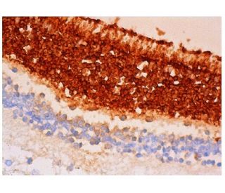
Visual Arrestin Antibody (G-2) is a mouse monoclonal IgG2a kappa light chain antibody developed to detect Visual Arrestin, also known as Arrestin 1, retinal S-antigen, or S-Arrestin, in human, mouse, and rat species through applications such as western blotting (WB), immunoprecipitation (IP), immunofluorescence (IF), immunohistochemistry with paraffin-embedded sections (IHCP), and enzyme-linked immunosorbent assay (ELISA). Visual Arrestin monoclonal antibody (G-2) is raised against amino acids 316-405 at the C-terminus of the human Visual Arrestin protein, ensuring effective binding for reliable detection. Visual Arrestin is primarily localized in the outer segments of retinal photoreceptor cells, where Visual Arrestin plays a crucial role in the phototransduction pathway by binding to phosphorylated rhodopsin. This interaction terminates the light signal, preventing continuous activation of G protein-coupled receptors and ensuring precise regulation of visual signals. Visual Arrestin is expressed in the pineal gland, where Visual Arrestin contributes to the regulation of circadian rhythms, highlighting Visual Arrestin′s importance in both vision and overall physiological processes. The strategic cellular localization of Visual Arrestin is essential for maintaining accurate visual perception and preventing signal desensitization, thereby enabling organisms to respond appropriately to changes in light conditions. Visual Arrestin serves as a major pathogenic autoantigen in inflammatory eye diseases such as uveoretinitis and Oguchi disease, a rare form of night blindness, underscoring Visual Arrestin′s significance in ocular health and disease. Anti-Visual Arrestin antibody (G-2) facilitates detailed studies of Visual Arrestin′s function and localization, aiding research into visual signal transduction, retinal disorders, and autoimmune conditions.
Ordering Information
| Product Name | Catalog # | UNIT | Price | Qty | FAVORITES | |
Visual Arrestin Antibody (G-2) | sc-166353 | 200 µg/ml | $322.00 | |||
Visual Arrestin Antibody (G-2): m-IgG Fc BP-HRP Bundle | sc-537478 | 200 µg Ab; 10 µg BP | $361.00 | |||
Visual Arrestin Antibody (G-2): m-IgGκ BP-HRP Bundle | sc-534757 | 200 µg Ab; 40 µg BP | $361.00 | |||
Visual Arrestin Antibody (G-2): m-IgG2a BP-HRP Bundle | sc-548026 | 200 µg Ab; 10 µg BP | $361.00 |