")
Visual Arrestin Antibody (F-2): sc-365029
- Visual Arrestin Antibody (F-2) is a mouse monoclonal IgG3 κ provided at 200 µg/ml
- specific for an epitope mapping between amino acids 339-371 near the C-terminus of Visual Arrestin of human origin
- recommended for detection of Visual Arrestin of mouse, rat and human origin by WB, IP, IF, IHC(P) and ELISA; also reactive with additional species, including and equine, canine, bovine and porcine
- m-IgGκ BP-HRP is the preferred secondary detection reagent for Visual Arrestin Antibody (F-2) for WB and IHC(P) applications. This reagent is now offered in a bundle with Visual Arrestin Antibody (F-2) (see ordering information below). For additional m-IgGκ BP conjugates see our complete list of Mouse IgG Binding Proteins.

QUICK LINKS
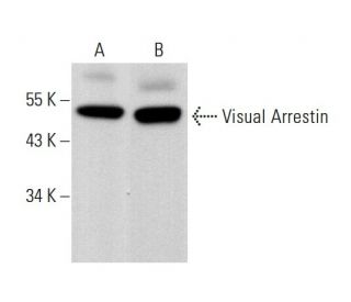
Visual Arrestin Antibody (F-2) is a mouse monoclonal IgG3 kappa light chain antibody developed to detect Visual Arrestin, also known as Arrestin 1, retinal S-antigen, or S-Arrestin, in multiple species including human, mouse, rat, equine, canine, bovine, and porcine. Visual Arrestin monoclonal antibody (F-2) targets an epitope within amino acids 339-371 near the C-terminus of the human Visual Arrestin protein, with detection in applications such as western blotting (WB), immunoprecipitation (IP), immunofluorescence (IF), immunohistochemistry with paraffin-embedded sections (IHCP), and enzyme-linked immunosorbent assay (ELISA). Visual Arrestin is predominantly localized in the outer segments of retinal photoreceptor cells and the pineal gland, where Visual Arrestin plays a crucial role in the phototransduction pathway by binding to phosphorylated rhodopsin. This binding effectively terminates the light signal, preventing continuous activation of G protein-coupled receptors and ensuring precise regulation of visual signals, which is essential for maintaining accurate visual perception and adapting to varying light conditions. Additionally, Visual Arrestin interacts with G protein-coupled receptor kinases (GRKs) to facilitate receptor desensitization and promote receptor internalization through clathrin-mediated endocytosis, further regulating visual signal transduction. The strategic cellular localization of Visual Arrestin in photoreceptor cells is vital for deactivating rhodopsin, thereby maintaining cellular homeostasis and proper visual function. anti-Visual Arrestin antibody (F-2) enables comprehensive studies of Visual Arrestin localization and function, supporting research into visual signal transduction, retinal disorders, and autoimmune conditions.
Ordering Information
| Product Name | Catalog # | UNIT | Price | Qty | FAVORITES | |
Visual Arrestin Antibody (F-2) | sc-365029 | 200 µg/ml | $322.00 | |||
Visual Arrestin Antibody (F-2): m-IgGκ BP-HRP Bundle | sc-522122 | 200 µg Ab, 40 µg BP | $361.00 | |||
Visual Arrestin (F-2) Neutralizing Peptide | sc-365029 P | 100 µg/0.5 ml | $69.00 |