")
Visual Arrestin Antibody (A-3): sc-271466
- Visual Arrestin Antibody (A-3) is a mouse monoclonal IgG2b κ, cited in 2 publications, provided at 200 µg/ml
- specific for an epitope mapping between amino acids 339-371 near the C-terminus of Visual Arrestin of human origin
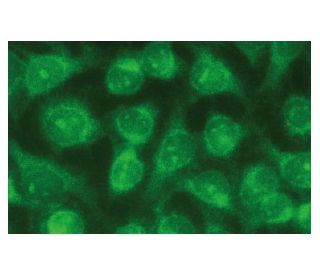
- recommended for detection of Visual Arrestin of mouse, rat and human origin by WB, IP, IF and ELISA; also reactive with additional species, including and equine, canine, bovine and porcine
- m-IgG2b BP-HRP is the preferred secondary detection reagent for Visual Arrestin Antibody (A-3) for WB applications. This reagent is now offered in a bundle with Visual Arrestin Antibody (A-3) (see ordering information below).

QUICK LINKS
Visual Arrestin Antibody (A-3) is a mouse monoclonal IgG2b kappa light chain antibody developed to detect Visual Arrestin, also known as Arrestin 1, retinal S-antigen, or S-Arrestin, across multiple species including human, mouse, rat, equine, canine, bovine, and porcine. Anti-Visual Arrestin antibody (A-3) targets an epitope within amino acids 339-371 near the C-terminus of the human Visual Arrestin protein, ensuring reliable detection in applications such as western blotting (WB), immunoprecipitation (IP), immunofluorescence (IF), immunohistochemistry with paraffin-embedded sections (IHCP), and enzyme-linked immunosorbent assay (ELISA). Visual Arrestin plays a fundamental role in the phototransduction pathway by binding to phosphorylated rhodopsin, thereby terminating the light signal and preventing the continuous activation of G protein-coupled receptors. This function is crucial for maintaining accurate visual perception and preventing signal desensitization, allowing organisms to adapt to varying light conditions effectively. Additionally, Visual Arrestin interacts with G protein-coupled receptor kinases (GRKs) to facilitate receptor desensitization and promote receptor internalization through clathrin-mediated endocytosis, further regulating visual signal transduction. Visual Arrestin is predominantly localized in the outer segments of retinal photoreceptor cells and the pineal gland, where Visual Arrestin not only contributes to vision but also plays a role in regulating circadian rhythms. The precise cellular localization and interaction with key proteins like rhodopsin and GRKs highlight the importance of Visual Arrestin in both visual and physiological processes. Moreover, Visual Arrestin serves as a major pathogenic autoantigen in inflammatory eye diseases such as uveoretinitis and Oguchi disease, a rare form of night blindness, underscoring its significance in ocular health and disease. Visual Arrestin (A-3) antibody facilitates comprehensive studies of this protein′s function and interactions, supporting research into visual signal transduction, retinal disorders, and autoimmune conditions.
Ordering Information
| Product Name | Catalog # | UNIT | Price | Qty | FAVORITES | |
Visual Arrestin Antibody (A-3) | sc-271466 | 200 µg/ml | $322.00 | |||
Visual Arrestin Antibody (A-3): m-IgG2b BP-HRP Bundle | sc-548540 | 200 µg Ab; 10 µg BP | $361.00 | |||
Visual Arrestin (A-3) Neutralizing Peptide | sc-271466 P | 100 µg/0.5 ml | $69.00 |