")
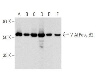
V-ATPase B2 Antibody (C-9): sc-166122
- V-ATPase B2 Antibody (C-9) is a mouse monoclonal IgG3 κ, cited in 4 publications, provided at 200 µg/ml
- specific for an epitope mapping between amino acids 2-38 at the N-terminus of V-ATPase B2 of human origin
- recommended for detection of V-ATPase B2 of mouse, rat and human origin by WB, IP, IF and ELISA; also reactive with additional species, including and canine and bovine
- m-IgG3 BP-HRP is the preferred secondary detection reagent for V-ATPase B2 Antibody (C-9) for WB applications. This reagent is now offered in a bundle with V-ATPase B2 Antibody (C-9) (see ordering information below).

QUICK LINKS
SEE ALSO...
V-ATPase B2 Antibody (C-9) is a mouse monoclonal IgG3 antibody that detects V-ATPase B2 in mouse, rat, and human samples through applications such as western blotting (WB), immunoprecipitation (IP), immunofluorescence (IF), and enzyme-linked immunosorbent assay (ELISA). V-ATPase B2 is a crucial component of the vacuolar-type H+-ATPase complex, which plays a vital role in maintaining cellular pH and ion homeostasis by facilitating proton transport across membranes. V-ATPase B2 is predominantly located in the kidney and osteoclasts, where V-ATPase B2 is essential for bone resorption and renal acidification. Proper functioning of V-ATPase B2 is critical for normal physiological processes, as V-ATPase B2 helps regulate the acidic environment necessary for various cellular functions, including enzyme activity and metabolic processes. Notably, mutations in the gene encoding V-ATPase B2 can lead to significant health issues, including impaired bone density and renal tubular acidosis. Anti-V-ATPase B2 antibody (C-9) provides a reliable tool for researchers investigating the role of V-ATPase B2 in health and disease, enabling a deeper understanding of its function and interactions within the cellular environment.
Alexa Fluor® is a trademark of Molecular Probes Inc., OR., USA
LI-COR® and Odyssey® are registered trademarks of LI-COR Biosciences
V-ATPase B2 Antibody (C-9) References:
- Distribution of Rab GTPases in mouse kidney and comparison with vacuolar H+-ATPase. | Curtis, LM. and Gluck, S. 2005. Nephron Physiol. 100: p31-42. PMID: 15838183
- Relocalization of the V-ATPase B2 subunit to the apical membrane of epididymal clear cells of mice deficient in the B1 subunit. | Da Silva, N., et al. 2007. Am J Physiol Cell Physiol. 293: C199-210. PMID: 17392376
- Compensatory membrane expression of the V-ATPase B2 subunit isoform in renal medullary intercalated cells of B1-deficient mice. | Paunescu, TG., et al. 2007. Am J Physiol Renal Physiol. 293: F1915-26. PMID: 17898041
- Role of CFTR and ClC-5 in modulating vacuolar H+-ATPase activity in kidney proximal tubule. | Carraro-Lacroix, LR., et al. 2010. Cell Physiol Biochem. 26: 563-76. PMID: 21063094
- V-ATPase promotes transforming growth factor-β-induced epithelial-mesenchymal transition of rat proximal tubular epithelial cells. | Cao, X., et al. 2012. Am J Physiol Renal Physiol. 302: F1121-32. PMID: 22129967
- Interaction between V-ATPase B2 and (Pro) renin Receptors in Promoting the progression of Renal Tubulointerstitial Fibrosis. | Liu, Y., et al. 2016. Sci Rep. 6: 25035. PMID: 27121029
- Impaired V-ATPase leads to increased lysosomal pH, results in disrupted lysosomal degradation and autophagic flux blockage, contributes to fluoride-induced developmental neurotoxicity. | Han, X., et al. 2022. Ecotoxicol Environ Saf. 236: 113500. PMID: 35421827
- Overexpression of V-ATPase B2 attenuates lung injury/fibrosis by stabilizing lysosomal membrane permeabilization and increasing collagen degradation. | Lee, JU., et al. 2022. Exp Mol Med. 54: 662-672. PMID: 35624153
- V-ATPase B2 promotes microglial phagocytosis of myelin debris by inactivating the MAPK signaling pathway. | Li, Y., et al. 2024. Neuropeptides. 106: 102436. PMID: 38733728
- F-53B disrupts energy metabolism by inhibiting the V-ATPase-AMPK axis in neuronal cells. | Zhang, Y., et al. 2025. J Hazard Mater. 487: 137111. PMID: 39793390
Ordering Information
| Product Name | Catalog # | UNIT | Price | Qty | FAVORITES | |
V-ATPase B2 Antibody (C-9) | sc-166122 | 200 µg/ml | $322.00 | |||
V-ATPase B2 Antibody (C-9): m-IgG3 BP-HRP Bundle | sc-550390 | 200 µg Ab; 40 µg BP | $361.00 | |||
V-ATPase B2 (C-9) Neutralizing Peptide | sc-166122 P | 100 µg/0.5 ml | $69.00 |