")
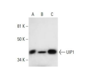
: sc-393259. Western blot analysis of UIP1 expression in NIH/3T3 (A), CSMLO (B) and F9 (C) whole cell lysates.")
: sc-393259. Western blot analysis of UIP1 expression in F9 (A) and U-251-MG (B) whole cell lysates.")
: sc-393259. Western blot analysis of UIP1 expression in non-transfected: sc-117752 (A) and mouse UIP1 transfected: sc-124465 (B) 293T whole cell lysates.")
UIP1 Antibody (C-2): sc-393259
- UIP1 Antibody (C-2) is a mouse monoclonal IgG1 κ UIP1 antibody provided at 200 µg/ml
- raised against amino acids 1-300 mapping at the N-terminus of UIP1 of mouse origin
- UIP1 Antibody (C-2) is recommended for detection of UIP1 of mouse, rat and human origin by WB, IP, IF and ELISA
- Anti-UIP1 Antibody (C-2) is available conjugated to agarose for IP; HRP for WB, IHC(P) and ELISA; and to either phycoerythrin or FITC for IF, IHC(P) and FCM
- also available conjugated to Alexa Fluor® 488, Alexa Fluor® 546, Alexa Fluor® 594 or Alexa Fluor® 647 for WB (RGB), IF, IHC(P) and FCM, and for use with RGB fluorescent imaging systems, such as iBright™ FL1000, FluorChem™, Typhoon, Azure and other comparable systems
- also available conjugated to Alexa Fluor® 680 or Alexa Fluor® 790 for WB (NIR), IF and FCM; for use with Near-Infrared (NIR) detection systems, such as LI-COR®Odyssey®, iBright™ FL1000, FluorChem™, Typhoon, Azure and other comparable systems
- m-IgG Fc BP-HRP and m-IgG1 BP-HRP are the preferred secondary detection reagents for UIP1 Antibody (C-2) for WB applications. These reagents are now offered in bundles with UIP1 Antibody (C-2) (see ordering information below).

QUICK LINKS
UIP1 Antibody (C-2) is a mouse monoclonal IgG1 kappa light chain antibody that detects UIP1 protein of mouse, rat, and human origin by western blotting (WB), immunoprecipitation (IP), immunofluorescence (IF), and enzyme-linked immunosorbent assay (ELISA). UIP1 (C-2) antibody is available in both non-conjugated and various conjugated forms, including agarose, horseradish peroxidase (HRP), phycoerythrin (PE), fluorescein isothiocyanate (FITC), and multiple Alexa Fluor® conjugates. UIP1, also known as UCHL5-interacting protein and HAUS augmin-like complex subunit 7, is a 368 amino acid protein encoded by the human HAUS7 gene. UIP1 plays a crucial role in regulating spindle assembly during mitosis, which is essential for accurate chromosome segregation and cell division. UIP1 functions as a key component of the HAUS augmin-like complex, which interacts with the gamma-tubulin ring complex to facilitate microtubule formation necessary for spindle assembly. Additionally, UIP1 binds to UCHL5, a protease that specifically cleaves ′Lys-48′-linked polyubiquitin chains, linking UIP1 to protein degradation pathways. UIP1 localizes to the centrosome during interphase and incorporates into spindle microtubules during mitosis, highlighting UIP1′s importance in maintaining centrosome integrity and proper mitotic function. Detectable UIP1 expression has been observed in various tissues, including the spleen, thymus, testis, ovary, small intestine, and colon, underscoring UIP1′s potential relevance in both normal physiology and disease states.
Alexa Fluor® is a trademark of Molecular Probes Inc., OR., USA
LI-COR® and Odyssey® are registered trademarks of LI-COR Biosciences
UIP1 Antibody (C-2) References:
- Identification of two proteins, S14 and UIP1, that interact with UCH37. | Li, T., et al. 2001. FEBS Lett. 488: 201-5. PMID: 11163772
- A quantitative atlas of mitotic phosphorylation. | Dephoure, N., et al. 2008. Proc Natl Acad Sci U S A. 105: 10762-7. PMID: 18669648
- Evaluation of the low-specificity protease elastase for large-scale phosphoproteome analysis. | Wang, B., et al. 2008. Anal Chem. 80: 9526-33. PMID: 19007248
- The augmin complex plays a critical role in spindle microtubule generation for mitotic progression and cytokinesis in human cells. | Uehara, R., et al. 2009. Proc Natl Acad Sci U S A. 106: 6998-7003. PMID: 19369198
- HAUS, the 8-subunit human Augmin complex, regulates centrosome and spindle integrity. | Lawo, S., et al. 2009. Curr Biol. 19: 816-26. PMID: 19427217
- Quantitative phosphoproteomic analysis of T cell receptor signaling reveals system-wide modulation of protein-protein interactions. | Mayya, V., et al. 2009. Sci Signal. 2: ra46. PMID: 19690332
- Expressed STSs and transcription of human Xq28. | Esposito, T., et al. 1997. Gene. 187: 185-91. PMID: 9099879
Ordering Information
| Product Name | Catalog # | UNIT | Price | Qty | FAVORITES | |
UIP1 Antibody (C-2) | sc-393259 | 200 µg/ml | $322.00 | |||
UIP1 Antibody (C-2): m-IgG Fc BP-HRP Bundle | sc-527675 | 200 µg Ab; 10 µg BP | $361.00 | |||
UIP1 Antibody (C-2): m-IgG1 BP-HRP Bundle | sc-533048 | 200 µg Ab; 20 µg BP | $361.00 | |||
UIP1 Antibody (C-2) AC | sc-393259 AC | 500 µg/ml, 25% agarose | $424.00 | |||
UIP1 Antibody (C-2) HRP | sc-393259 HRP | 200 µg/ml | $322.00 | |||
UIP1 Antibody (C-2) FITC | sc-393259 FITC | 200 µg/ml | $336.00 | |||
UIP1 Antibody (C-2) PE | sc-393259 PE | 200 µg/ml | $349.00 | |||
UIP1 Antibody (C-2) Alexa Fluor® 488 | sc-393259 AF488 | 200 µg/ml | $364.00 | |||
UIP1 Antibody (C-2) Alexa Fluor® 546 | sc-393259 AF546 | 200 µg/ml | $364.00 | |||
UIP1 Antibody (C-2) Alexa Fluor® 594 | sc-393259 AF594 | 200 µg/ml | $364.00 | |||
UIP1 Antibody (C-2) Alexa Fluor® 647 | sc-393259 AF647 | 200 µg/ml | $364.00 | |||
UIP1 Antibody (C-2) Alexa Fluor® 680 | sc-393259 AF680 | 200 µg/ml | $364.00 | |||
UIP1 Antibody (C-2) Alexa Fluor® 790 | sc-393259 AF790 | 200 µg/ml | $364.00 |