")
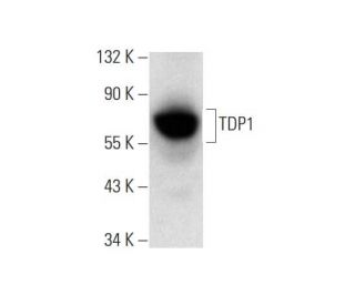
: sc-365674. Western blot analysis of TDP1 expression in Ramos whole cell lysate.")
: sc-365674. Immunoperoxidase staining of formalin fixed, paraffin-embedded human thyroid gland tissue showing cytoplasmic staining of glandular cells.")
: sc-365674. Near-Infrared western blot analysis of TDP1 expression in Ramos whole cell lysate. Blocked with UltraCruz<sup>®</sup> Blocking Reagent: sc-516214. Detection reagent used: m-IgGκ BP-CFL 647: sc-516179.")
TDP1 Antibody (C-3): sc-365674
- TDP1 Antibody (C-3) is a mouse monoclonal IgG1 κ TDP1 antibody, cited in 10 publications, provided at 200 µg/ml
- raised against amino acids 309-608 mapping at the C-terminus of TDP1 of human origin
- TDP1 Antibody (C-3) is recommended for detection of TDP1 of human origin by WB, IP, IF, IHC(P) and ELISA
- Anti-TDP1 Antibody (C-3) is available conjugated to agarose for IP; HRP for WB, IHC(P) and ELISA; and to either phycoerythrin or FITC for IF, IHC(P) and FCM
- also available conjugated to Alexa Fluor® 488, Alexa Fluor® 546, Alexa Fluor® 594 or Alexa Fluor® 647 for WB (RGB), IF, IHC(P) and FCM, and for use with RGB fluorescent imaging systems, such as iBright™ FL1000, FluorChem™, Typhoon, Azure and other comparable systems
- also available conjugated to Alexa Fluor® 680 or Alexa Fluor® 790 for WB (NIR), IF and FCM; for use with Near-Infrared (NIR) detection systems, such as LI-COR®Odyssey®, iBright™ FL1000, FluorChem™, Typhoon, Azure and other comparable systems
- m-IgG Fc BP-HRP and m-IgG1 BP-HRP are the preferred secondary detection reagents for TDP1 Antibody (C-3) for WB and IHC(P) applications. These reagents are now offered in bundles with TDP1 Antibody (C-3) (see ordering information below).

QUICK LINKS
SEE ALSO...
TDP1 Antibody (C-3) is a mouse monoclonal IgG1 kappa light chain antibody that detects TDP1 protein of human origin by western blotting (WB), immunoprecipitation (IP), immunofluorescence (IF), immunohistochemistry, and enzyme-linked immunosorbent assay (ELISA). TDP1 (C-3) antibody is available in both non-conjugated and various conjugated forms, including agarose, horseradish peroxidase (HRP), phycoerythrin (PE), fluorescein isothiocyanate (FITC), and multiple Alexa Fluor® conjugates. Tyrosyl-DNA phosphodiesterase 1 (TDP1) plays a crucial role in DNA repair by catalyzing the hydrolysis of phosphodiester bonds between tyrosine residues and DNA 3′-phosphates, which is essential for maintaining genomic stability. TDP1 is particularly important in the repair of DNA damage caused by free radicals, as TDP1 also removes glycolate from single-stranded DNA containing a 3′-phosphoglycolate, thereby facilitating the repair of double-strand breaks. The presence of a unique HKD signature motif, characterized by highly conserved lysine and histidine residues, classifies TDP1 within the phospholipase D superfamily, highlighting evolutionary significance. The hydrolytic reaction catalyzed by TDP1 involves a phosphoryl transfer mechanism that is common to all members of this superfamily. Loss-of-function mutations in TDP1 can lead to severe neurological disorders, such as spinocerebellar ataxia with axonal neuropathy, by disrupting DNA transcription and inducing apoptosis in postmitotic neurons, underscoring TDP1′s vital role in cellular health and function.
Alexa Fluor® is a trademark of Molecular Probes Inc., OR., USA
LI-COR® and Odyssey® are registered trademarks of LI-COR Biosciences
TDP1 Antibody (C-3) References:
- The tyrosyl-DNA phosphodiesterase Tdp1 is a member of the phospholipase D superfamily. | Interthal, H., et al. 2001. Proc Natl Acad Sci U S A. 98: 12009-14. PMID: 11572945
- Conversion of phosphoglycolate to phosphate termini on 3' overhangs of DNA double strand breaks by the human tyrosyl-DNA phosphodiesterase hTdp1. | Inamdar, KV., et al. 2002. J Biol Chem. 277: 27162-8. PMID: 12023295
- Mutation of TDP1, encoding a topoisomerase I-dependent DNA damage repair enzyme, in spinocerebellar ataxia with axonal neuropathy. | Takashima, H., et al. 2002. Nat Genet. 32: 267-72. PMID: 12244316
- Insights into substrate binding and catalytic mechanism of human tyrosyl-DNA phosphodiesterase (Tdp1) from vanadate and tungstate-inhibited structures. | Davies, DR., et al. 2002. J Mol Biol. 324: 917-32. PMID: 12470949
Ordering Information
| Product Name | Catalog # | UNIT | Price | Qty | FAVORITES | |
TDP1 Antibody (C-3) | sc-365674 | 200 µg/ml | $322.00 | |||
TDP1 Antibody (C-3): m-IgG Fc BP-HRP Bundle | sc-527389 | 200 µg Ab; 10 µg BP | $361.00 | |||
TDP1 Antibody (C-3): m-IgG1 BP-HRP Bundle | sc-532762 | 200 µg Ab; 20 µg BP | $361.00 | |||
TDP1 Antibody (C-3) AC | sc-365674 AC | 500 µg/ml, 25% agarose | $424.00 | |||
TDP1 Antibody (C-3) HRP | sc-365674 HRP | 200 µg/ml | $322.00 | |||
TDP1 Antibody (C-3) FITC | sc-365674 FITC | 200 µg/ml | $336.00 | |||
TDP1 Antibody (C-3) PE | sc-365674 PE | 200 µg/ml | $349.00 | |||
TDP1 Antibody (C-3) Alexa Fluor® 488 | sc-365674 AF488 | 200 µg/ml | $364.00 | |||
TDP1 Antibody (C-3) Alexa Fluor® 546 | sc-365674 AF546 | 200 µg/ml | $364.00 | |||
TDP1 Antibody (C-3) Alexa Fluor® 594 | sc-365674 AF594 | 200 µg/ml | $364.00 | |||
TDP1 Antibody (C-3) Alexa Fluor® 647 | sc-365674 AF647 | 200 µg/ml | $364.00 | |||
TDP1 Antibody (C-3) Alexa Fluor® 680 | sc-365674 AF680 | 200 µg/ml | $364.00 | |||
TDP1 Antibody (C-3) Alexa Fluor® 790 | sc-365674 AF790 | 200 µg/ml | $364.00 |