")
SCF Antibody (G-3): sc-13126
- SCF Antibody (G-3) is a mouse monoclonal IgG2b κ SCF antibody, cited in 53 publications, provided at 200 µg/ml
- raised against amino acids 26-214 of SCF of human origin
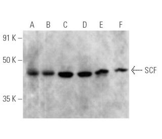
- SCF Antibody (G-3) is recommended for detection of SCF of mouse, rat and human origin by WB, IP, IF, IHC(P) and ELISA
- Anti-SCF Antibody (G-3) is available conjugated to agarose for IP; HRP for WB, IHC(P) and ELISA; and to either phycoerythrin or FITC for IF, IHC(P) and FCM
- also available conjugated to Alexa Fluor® 488, Alexa Fluor® 546, Alexa Fluor® 594 or Alexa Fluor® 647 for WB (RGB), IF, IHC(P) and FCM, and for use with RGB fluorescent imaging systems, such as iBright™ FL1000, FluorChem™, Typhoon, Azure and other comparable systems
- also available conjugated to Alexa Fluor® 680 or Alexa Fluor® 790 for WB (NIR), IF and FCM; for use with Near-Infrared (NIR) detection systems, such as LI-COR®Odyssey®, iBright™ FL1000, FluorChem™, Typhoon, Azure and other comparable systems
- m-IgG2b BP-HRP and m-IgGκ BP-HRP are the preferred secondary detection reagents for SCF Antibody (G-3) for WB and IHC(P) applications. These reagents are now offered in bundles with SCF Antibody (G-3) (see ordering information below).

QUICK LINKS
SEE ALSO...
SCF Antibody (G-3) is a mouse monoclonal IgG2b kappa light chain antibody that detects SCF protein of mouse, rat, and human origin by WB, IP, IF, IHCP, and ELISA. SCF Antibody (G-3) is available in both non-conjugated and various conjugated forms, including agarose, HRP, PE, FITC, and multiple Alexa Fluor® conjugates. SCF plays a crucial role as the ligand for the transmembrane tyrosine kinase receptor proto-oncogene c-Kit, which is vital for several cellular processes. SCF is a pleiotropic cytokine that exists in two alternatively spliced forms, consisting of 248 and 220 amino acids in human and mouse systems, respectively. The larger form of SCF is predominantly expressed in fibroblasts, brain, and thymus, while the smaller variant is found in tissues such as the spleen, testis, placenta, and cerebellum. SCF is essential for the development of germ cells, hematopoietic progenitor cells, and melanocyte precursors, as SCF stimulates the proliferation and maturation of both mature and immature mast cells. The interaction of SCF with c-Kit is critical for various biological processes, including hematopoiesis and pigmentation, making SCF monoclonal antibody (G-3) a valuable tool for research in developmental biology and cancer studies.
Alexa Fluor® is a trademark of Molecular Probes Inc., OR., USA
LI-COR® and Odyssey® are registered trademarks of LI-COR Biosciences
SCF Antibody (G-3) References:
- Differential expression and processing of two cell associated forms of the kit-ligand: KL-1 and KL-2. | Huang, EJ., et al. 1992. Mol Biol Cell. 3: 349-62. PMID: 1378327
- Support of human hematopoiesis in long-term bone marrow cultures by murine stromal cells selectively expressing the membrane-bound and secreted forms of the human homolog of the steel gene product, stem cell factor. | Toksoz, D., et al. 1992. Proc Natl Acad Sci U S A. 89: 7350-4. PMID: 1380155
- Cleavage of membrane-anchored growth factors involves distinct protease activities regulated through common mechanisms. | Pandiella, A., et al. 1992. J Biol Chem. 267: 24028-33. PMID: 1385433
- Mast cell growth factor maps near the steel locus on mouse chromosome 10 and is deleted in a number of steel alleles. | Copeland, NG., et al. 1990. Cell. 63: 175-83. PMID: 1698554
- Primary structure and functional expression of rat and human stem cell factor DNAs. | Martin, FH., et al. 1990. Cell. 63: 203-11. PMID: 2208279
- Role of stem cell factor in the placental niche. | Khodadi, E., et al. 2016. Cell Tissue Res. 366: 523-531. PMID: 27234501
- Erythropoietin, Stem Cell Factor, and Cancer Cell Migration. | Vazquez-Mellado, MJ., et al. 2017. Vitam Horm. 105: 273-296. PMID: 28629522
- Bone marrow adipocytes promote the regeneration of stem cells and haematopoiesis by secreting SCF. | Zhou, BO., et al. 2017. Nat Cell Biol. 19: 891-903. PMID: 28714970
- Stem cell factor: the bridge between bone marrow adipocytes and hematopoietic cells. | Li, Z. and MacDougald, OA. 2019. Haematologica. 104: 1689-1691. PMID: 31473604
- Inhibition of Soluble Stem Cell Factor Promotes Intestinal Mucosal Repair. | Garcia-Hernandez, V., et al. 2023. Inflamm Bowel Dis. 29: 1133-1144. PMID: 36688460
- Stem cell factor and cKIT modulate endothelial glycolysis in hypoxia. | Jeong, H., et al. 2024. Cardiovasc Res. 120: 745-755. PMID: 38507654
- Monocytes do not make mast cells when cultured in the presence of SCF. Characterization of the circulating mast cell progenitor as a c-kit+, CD34+, Ly-, CD14-, CD17-, colony-forming cell. | Agis, H., et al. 1993. J Immunol. 151: 4221-7. PMID: 7691941
Ordering Information
| Product Name | Catalog # | UNIT | Price | Qty | FAVORITES | |
SCF Antibody (G-3) | sc-13126 | 200 µg/ml | $322.00 | |||
SCF Antibody (G-3): m-IgGκ BP-HRP Bundle | sc-520598 | 200 µg Ab, 40 µg BP | $361.00 | |||
SCF Antibody (G-3): m-IgG2b BP-HRP Bundle | sc-548841 | 200 µg Ab; 10 µg BP | $361.00 | |||
SCF Antibody (G-3) AC | sc-13126 AC | 500 µg/ml, 25% agarose | $424.00 | |||
SCF Antibody (G-3) HRP | sc-13126 HRP | 200 µg/ml | $322.00 | |||
SCF Antibody (G-3) FITC | sc-13126 FITC | 200 µg/ml | $336.00 | |||
SCF Antibody (G-3) PE | sc-13126 PE | 200 µg/ml | $349.00 | |||
SCF Antibody (G-3) Alexa Fluor® 488 | sc-13126 AF488 | 200 µg/ml | $364.00 | |||
SCF Antibody (G-3) Alexa Fluor® 546 | sc-13126 AF546 | 200 µg/ml | $364.00 | |||
SCF Antibody (G-3) Alexa Fluor® 594 | sc-13126 AF594 | 200 µg/ml | $364.00 | |||
SCF Antibody (G-3) Alexa Fluor® 647 | sc-13126 AF647 | 200 µg/ml | $364.00 | |||
SCF Antibody (G-3) Alexa Fluor® 680 | sc-13126 AF680 | 200 µg/ml | $364.00 | |||
SCF Antibody (G-3) Alexa Fluor® 790 | sc-13126 AF790 | 200 µg/ml | $364.00 |