")
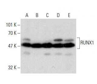
RUNX1 Antibody (DW71): sc-101146
- RUNX1 Antibody (DW71) is a mouse monoclonal IgG2b κ, cited in 15 publications, provided at 100 µg/ml
- raised against recombinant RUNX1 of human origin
- recommended for detection of a broad range of RUNX1 isoforms of mouse, rat and human origin by WB, IP, IF, IHC(P) and ELISA
- See RUNX1 (A-2): sc-365644 for RUNX1 antibody conjugates, including AC, HRP, FITC, PE, Alexa Fluor® 488, 594, 647, 680 and 790.
- At present, we have not yet completed the identification of the preferred secondary detection reagent(s) for RUNX1 Antibody (DW71). This work is in progress.
QUICK LINKS
RUNX1 Antibody (DW71) is a mouse monoclonal IgG2b kappa light chain antibody that detects RUNX1 protein of mouse, rat, and human origin by western blotting (WB), immunoprecipitation (IP), immunofluorescence (IF), immunohistochemistry with paraffin-embedded sections (IHCP), and enzyme-linked immunosorbent assay (ELISA). RUNX1 (DW71) antibody is available in a non-conjugated form, allowing for versatile applications in various experimental settings. RUNX1, also known as AML-1, PEBP2αB, or CBFA2, is a crucial member of the Runt-related transcription factor family, which includes RUNX2 and RUNX3. RUNX1 plays a vital role in hematopoiesis, the process of blood cell formation, and is frequently implicated in human leukemia due to chromosomal translocations that disrupt function by fusing with other transcription factors and corepressor molecules. Additionally, RUNX1 is essential for sensory neuron diversification, promoting axonal growth and being selectively expressed in neural crest-derived TrkA+ sensory neurons, facilitating TrkA transactivation in migratory neural crest cells. RUNX1′s ability to regulate gene expression through alternative splicing results in several isoforms, each potentially contributing to diverse biological functions. Understanding RUNX1′s role in these processes is critical for elucidating involvement in both normal development and disease states, making anti-RUNX1 antibody (DW71) an invaluable tool for researchers studying these pathways.
Ordering Information
| Product Name | Catalog # | UNIT | Price | Qty | FAVORITES | |
RUNX1 Antibody (DW71) | sc-101146 | 100 µg/ml | $333.00 |