")
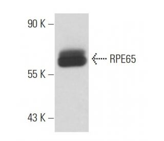
: sc-73616. Western blot analysis of RPE65 expression in mouse eye tissue extract.")
RPE65 Antibody (3D9): sc-73616
- RPE65 Antibody (3D9) is a mouse monoclonal IgG1 κ, cited in 4 publications, provided at 200 µg/ml
- raised against RPE microsomal membranes of bovine origin
- recommended for detection of RPE65 of mouse, rat, human and Xenopus origin by WB, IP and IF; also reactive with additional species, including bovine
- m-IgG Fc BP-HRP and m-IgG1 BP-HRP are the preferred secondary detection reagents for RPE65 Antibody (3D9) for WB applications. These reagents are now offered in bundles with RPE65 Antibody (3D9) (see ordering information below).

QUICK LINKS
SEE ALSO...
RPE65 Antibody (3D9) is a mouse monoclonal IgG1 antibody that detects RPE65 in mouse, rat, and human samples through applications such as western blotting (WB), immunoprecipitation (IP), and immunofluorescence (IF). RPE65 is a crucial protein located in the retinal pigment epithelium, where RPE65 plays a vital role in the visual cycle by facilitating the conversion of all-trans-retinal to 11-cis-retinal, a key component in the regeneration of visual pigments. This process is essential for maintaining photoreceptor function and overall vision. RPE65 exists in two forms: a membrane-bound form that is palmitoylated, allowing RPE65 to anchor to the cell membrane, and a soluble form that binds vitamin A when unpalmitoylated. Deficiencies or mutations in RPE65 can lead to severe visual impairments, including autosomal dominant retinitis pigmentosa and Leber congenital amaurosis type 2, highlighting the importance of RPE65 in ocular health. Anti-RPE65 antibody (3D9) is an invaluable tool for researchers studying retinal diseases and the underlying mechanisms of vision restoration.
Alexa Fluor® is a trademark of Molecular Probes Inc., OR., USA
LI-COR® and Odyssey® are registered trademarks of LI-COR Biosciences
RPE65 Antibody (3D9) References:
- Function of the protein RPE65 in the visual cycle. | Wolf, G. 2005. Nutr Rev. 63: 97-100. PMID: 15825812
- The Consequences of Hypomorphic RPE65 for Rod and Cone Photoreceptors. | Samardzija, M., et al. 2016. Adv Exp Med Biol. 854: 341-6. PMID: 26427430
- Prospect of retinal gene therapy following commercialization of voretigene neparvovec-rzyl for retinal dystrophy mediated by RPE65 mutation. | Ameri, H. 2018. J Curr Ophthalmol. 30: 1-2. PMID: 29564403
- The effect of human gene therapy for RPE65-associated Leber's congenital amaurosis on visual function: a systematic review and meta-analysis. | Wang, X., et al. 2020. Orphanet J Rare Dis. 15: 49. PMID: 32059734
- Voretigene neparvovec-rzyl for treatment of RPE65-mediated inherited retinal diseases: a model for ocular gene therapy development. | Ciulla, TA., et al. 2020. Expert Opin Biol Ther. 20: 565-578. PMID: 32149547
- Epidemiology of Mutations in the 65-kDa Retinal Pigment Epithelium (RPE65) Gene-Mediated Inherited Retinal Dystrophies: A Systematic Literature Review. | Sallum, JMF., et al. 2022. Adv Ther. 39: 1179-1198. PMID: 35098484
- Frequency of RPE65 Gene Mutation in Patients with Hereditary Retinal Dystrophy. | Kahraman, NS., et al. 2022. Turk J Ophthalmol. 52: 270-275. PMID: 36017377
- The novel visual cycle inhibitor (±)-RPE65-61 protects retinal photoreceptors from light-induced degeneration. | Wang, Y., et al. 2022. PLoS One. 17: e0269437. PMID: 36227868
- Single Center Experience with Voretigene Neparvovec Gene Augmentation Therapy in RPE65 Mutation-Associated Inherited Retinal Degeneration in a Clinical Setting. | Lorenz, B., et al. 2024. Ophthalmology. 131: 161-178. PMID: 37704110
Ordering Information
| Product Name | Catalog # | UNIT | Price | Qty | FAVORITES | |
RPE65 Antibody (3D9) | sc-73616 | 200 µg/ml | $322.00 | |||
RPE65 Antibody (3D9): m-IgG Fc BP-HRP Bundle | sc-539503 | 200 µg Ab; 10 µg BP | $361.00 | |||
RPE65 Antibody (3D9): m-IgG1 BP-HRP Bundle | sc-541529 | 200 µg Ab; 20 µg BP | $361.00 |