")
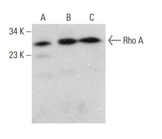
RhoA Antibody (F-1): sc-166399
- RhoA Antibody (F-1) is a mouse monoclonal IgG2b κ, cited in 8 publications, provided at 200 µg/ml
- specific for an epitope mapping between amino acids 116-140 within an internal region of Rho A of human origin
- recommended for detection of Rho A p21 and Rho C of mouse, rat, human and origin by WB, IP, IF, IHC(P) and ELISA; also reactive with additional species, including and equine, canine, bovine, porcine and avian
- TransCruz reagent for ChIP application (sc-166399 X, 200 µg/0.1 ml)
- See RhoA (26C4): sc-418 for RhoA antibody conjugates, including AC, HRP, FITC, PE, Alexa Fluor® 488, 594, 647, 680 and 790.
- m-IgG Fc BP-HRP and m-IgG2b BP-HRP are the preferred secondary detection reagents for RhoA Antibody (F-1) for WB and IHC(P) applications. These reagents are now offered in bundles with RhoA Antibody (F-1) (see ordering information below).

QUICK LINKS
SEE ALSO...
Rho A Antibody (F-1) is a mouse monoclonal IgG2b antibody that detects Rho A in mouse, rat, and human samples through various applications including western blotting (WB), immunoprecipitation (IP), immunofluorescence (IF), immunohistochemistry with paraffin-embedded sections (IHCP), and enzyme-linked immunosorbent assay (ELISA). Rho A is a member of the Rho family of GTPases, which play a crucial role in regulating the actin cytoskeleton, thereby influencing cell shape, motility, and adhesion. Rho A is primarily located in the cytoplasm and is activated by various extracellular signals, leading to translocation to the plasma membrane where Rho A interacts with downstream effectors. Proper localization of Rho A is vital for function in cellular processes such as migration and proliferation, which are essential for normal development and tissue homeostasis. Dysregulation of Rho A has been implicated in various pathological conditions, including cancer metastasis and cardiovascular diseases, making Rho A a significant target for therapeutic intervention. Anti-Rho A antibody (F-1) provides a reliable tool for researchers studying these critical pathways and their implications in health and disease.
Ordering Information
| Product Name | Catalog # | UNIT | Price | Qty | FAVORITES | |
RhoA Antibody (F-1) | sc-166399 | 200 µg/ml | $322.00 | |||
RhoA Antibody (F-1): m-IgG Fc BP-HRP Bundle | sc-540097 | 200 µg Ab; 10 µg BP | $361.00 | |||
RhoA Antibody (F-1): m-IgG2b BP-HRP Bundle | sc-549773 | 200 µg Ab; 10 µg BP | $361.00 | |||
RhoA (F-1) Neutralizing Peptide | sc-166399 P | 100 µg/0.5 ml | $69.00 | |||
RhoA Antibody (F-1) X | sc-166399 X | 200 µg/0.1 ml | $322.00 |