")
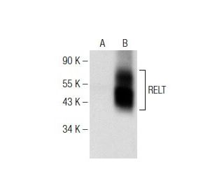
: sc-373942. Western blot analysis of RELT expression in non-transfected: sc-117752 (A) and human RELT transfected: sc-159793 (B) 293T whole cell lysates.")
: sc-373942. Western blot analysis of RELT expression in non-transfected 293T: sc-117752 (A), human RELT transfected 293T: sc-159793 (B), HeLa (C) and Jurkat (D) whole cell lysates.")
: sc-373942. Western blot analysis of RELT expression in HEL 92.1.7 whole cell lysate.")
RELT Antibody (C-6): sc-373942
- RELT Antibody (C-6) is a mouse monoclonal IgG1 κ RELT antibody, cited in 1 publications, provided at 200 µg/ml
- raised against amino acids 129-348 mapping within an internal region of RELT of human origin
- RELT Antibody (C-6) is recommended for detection of RELT of human origin by WB, IP, IF and ELISA
- Anti-RELT Antibody (C-6) is available conjugated to agarose for IP; HRP for WB, IHC(P) and ELISA; and to either phycoerythrin or FITC for IF, IHC(P) and FCM
- also available conjugated to Alexa Fluor® 488, Alexa Fluor® 546, Alexa Fluor® 594 or Alexa Fluor® 647 for WB (RGB), IF, IHC(P) and FCM, and for use with RGB fluorescent imaging systems, such as iBright™ FL1000, FluorChem™, Typhoon, Azure and other comparable systems
- also available conjugated to Alexa Fluor® 680 or Alexa Fluor® 790 for WB (NIR), IF and FCM; for use with Near-Infrared (NIR) detection systems, such as LI-COR®Odyssey®, iBright™ FL1000, FluorChem™, Typhoon, Azure and other comparable systems
- m-IgG Fc BP-HRP is the preferred secondary detection reagent for RELT Antibody (C-6) for WB applications. This reagent is now offered in a bundle with RELT Antibody (C-6) (see ordering information below).

QUICK LINKS
SEE ALSO...
RELT Antibody (C-6) is a mouse monoclonal IgG1 kappa light chain antibody that detects RELT protein of human origin by western blotting (WB), immunoprecipitation (IP), immunofluorescence (IF), and enzyme-linked immunosorbent assay (ELISA). RELT (C-6) antibody is available in both non-conjugated and various conjugated forms, including agarose, horseradish peroxidase (HRP), phycoerythrin (PE), fluorescein isothiocyanate (FITC), and multiple Alexa Fluor® conjugates. RELT protein, also known as receptor expressed in lymphoid tissues or tumor necrosis factor receptor superfamily member 19L (TNFRSF19L), is a transmembrane glycoprotein predominantly expressed in thymus, spleen, testis, colon, skeletal muscle, and peripheral blood lymphocytes. RELT′s unique structure includes two cysteine-rich domains, although one is incomplete, and notably lacks the death domain found in some other tumor necrosis factor receptor family members. This absence of the death domain differentiates RELT from other family members that can bind TRAF adaptor proteins, indicating a distinct signaling pathway. RELT interacts with and becomes phosphorylated by SPAK and OSR1 kinases, which are crucial for the activation of p38 and JNK signaling pathways. These interactions suggest RELT plays a vital role in T-cell activation, as RELT overexpression leads to phosphorylation of c-Jun and ATF2, further implicating RELT in the regulation of important cellular responses.
Alexa Fluor® is a trademark of Molecular Probes Inc., OR., USA
LI-COR® and Odyssey® are registered trademarks of LI-COR Biosciences
RELT Antibody (C-6) References:
- RELT, a new member of the tumor necrosis factor receptor superfamily, is selectively expressed in hematopoietic tissues and activates transcription factor NF-kappaB. | Sica, GL., et al. 2001. Blood. 97: 2702-7. PMID: 11313261
- Cytokine-receptor pairing: accelerating discovery of cytokine function. | Foster, D., et al. 2004. Nat Rev Drug Discov. 3: 160-70. PMID: 15040579
- Tumor necrosis factor family ligand-receptor binding. | Zhang, G. 2004. Curr Opin Struct Biol. 14: 154-60. PMID: 15093829
- Identification of RELT homologues that associate with RELT and are phosphorylated by OSR1. | Cusick, JK., et al. 2006. Biochem Biophys Res Commun. 340: 535-43. PMID: 16389068
- The TNF receptor, RELT, binds SPAK and uses it to mediate p38 and JNK activation. | Polek, TC., et al. 2006. Biochem Biophys Res Commun. 343: 125-34. PMID: 16530727
- Interactions of tumor necrosis factor (TNF) and TNF receptor family members in the mouse and human. | Bossen, C., et al. 2006. J Biol Chem. 281: 13964-71. PMID: 16547002
- TRAIL-induced cleavage and inactivation of SPAK sensitizes cells to apoptosis. | Polek, TC., et al. 2006. Biochem Biophys Res Commun. 349: 1016-24. PMID: 16950202
Ordering Information
| Product Name | Catalog # | UNIT | Price | Qty | FAVORITES | |
RELT Antibody (C-6) | sc-373942 | 200 µg/ml | $322.00 | |||
RELT Antibody (C-6): m-IgG Fc BP-HRP Bundle | sc-525930 | 200 µg Ab; 10 µg BP | $361.00 | |||
RELT Antibody (C-6) AC | sc-373942 AC | 500 µg/ml, 25% agarose | $424.00 | |||
RELT Antibody (C-6) HRP | sc-373942 HRP | 200 µg/ml | $322.00 | |||
RELT Antibody (C-6) FITC | sc-373942 FITC | 200 µg/ml | $336.00 | |||
RELT Antibody (C-6) PE | sc-373942 PE | 200 µg/ml | $349.00 | |||
RELT Antibody (C-6) Alexa Fluor® 488 | sc-373942 AF488 | 200 µg/ml | $364.00 | |||
RELT Antibody (C-6) Alexa Fluor® 546 | sc-373942 AF546 | 200 µg/ml | $364.00 | |||
RELT Antibody (C-6) Alexa Fluor® 594 | sc-373942 AF594 | 200 µg/ml | $364.00 | |||
RELT Antibody (C-6) Alexa Fluor® 647 | sc-373942 AF647 | 200 µg/ml | $364.00 | |||
RELT Antibody (C-6) Alexa Fluor® 680 | sc-373942 AF680 | 200 µg/ml | $364.00 | |||
RELT Antibody (C-6) Alexa Fluor® 790 | sc-373942 AF790 | 200 µg/ml | $364.00 |