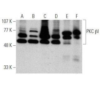
Date published: 2025-11-22

1-800-457-3801

SCBT Portrait Logo
Seach Input

PKC beta 1 Antibody (E-3): sc-8049

4.6(14)
Write a reviewAsk a question

Datasheets
  • PKC beta 1 Antibody (E-3) is a mouse monoclonal IgG1 κ PKC beta 1 antibody, cited in 51 publications, provided at 200 µg/ml
  • specific for an epitope mapping between amino acids 656-671 at the C-terminus of PKC βI of human origin
  • PKC beta 1 Antibody (E-3) is recommended for detection of PKC βI of mouse, rat, human and origin by WB, IP, IF, IHC(P) and ELISA; also reactive with additional species, including and canine, bovine, porcine and avian
  • Anti-PKC beta 1 Antibody (E-3) is available conjugated to agarose for IP; HRP for WB, IHC(P) and ELISA; and to either phycoerythrin or FITC for IF, IHC(P) and FCM
  • also available conjugated to Alexa Fluor® 488, Alexa Fluor® 546, Alexa Fluor® 594 or Alexa Fluor® 647 for WB (RGB), IF, IHC(P) and FCM, and for use with RGB fluorescent imaging systems, such as iBright™ FL1000, FluorChem™, Typhoon, Azure and other comparable systems
  • also available conjugated to Alexa Fluor® 680 or Alexa Fluor® 790 for WB (NIR), IF and FCM; for use with Near-Infrared (NIR) detection systems, such as LI-COR®Odyssey®, iBright™ FL1000, FluorChem™, Typhoon, Azure and other comparable systems
  • m-IgG Fc BP-HRP and m-IgGκ BP-HRP are the preferred secondary detection reagents for PKC beta 1 Antibody (E-3) for WB and IHC(P) applications. These reagents are now offered in bundles with PKC beta 1 Antibody (E-3) (see ordering information below).

QUICK LINKS

SEE ALSO...

PKC βI Antibody (E-3) is a mouse monoclonal IgG1 kappa light chain antibody that detects PKC beta I protein of mouse, rat, and human origin by western blotting (WB), immunoprecipitation (IP), immunofluorescence (IF), immunohistochemistry with paraffin embedded sections (IHCP), and enzyme-linked immunosorbent assay (ELISA). PKC βI (E-3) antibody is available in both non-conjugated and various conjugated forms, including agarose, horseradish peroxidase (HRP), phycoerythrin (PE), fluorescein isothiocyanate (FITC), and multiple Alexa Fluor® conjugates. PKC beta I protein, also known as PRKCB, is a crucial member of the protein kinase C (PKC) family, which plays a significant role in regulating diverse cellular functions such as cell growth, differentiation, gene expression, and hormone secretion. PKC beta I is primarily located in the cytoplasm and translocates to the membrane upon activation, which is essential for its role in signal transduction pathways. This translocation allows PKC beta I to interact with various substrates and participate in critical cellular processes, including the modulation of cell survival and apoptosis. Activation of PKC beta I is influenced by diacylglycerols and phorbol esters, which bind to PKC beta I and promote kinase activity. Understanding PKC beta I localization and function is important for elucidating involvement in various diseases, including cancer and cardiovascular disorders, making PKC βI (E-3) monoclonal antibody an invaluable tool for researchers studying these pathways.

For Research Use Only. Not Intended for Diagnostic or Therapeutic Use.

Alexa Fluor® is a trademark of Molecular Probes Inc., OR., USA

LI-COR® and Odyssey® are registered trademarks of LI-COR Biosciences

PKC beta 1 Antibody (E-3) References:

  1. A new member of the protein kinase C family, nPKC theta, predominantly expressed in skeletal muscle.  |  Osada, S., et al. 1992. Mol Cell Biol. 12: 3930-8. PMID: 1508194
  2. Structural and functional diversities of a family of signal transducing protein kinases, protein kinase C family; two distinct classes of PKC, conventional cPKC and novel nPKC.  |  Ohno, S., et al. 1991. Adv Enzyme Regul. 31: 287-303. PMID: 1877391
  3. Expression and characterization of protein kinase C-delta.  |  Olivier, AR. and Parker, PJ. 1991. Eur J Biochem. 200: 805-10. PMID: 1915352
  4. Calcium-dependent activation of a multifunctional protein kinase by membrane phospholipids.  |  Takai, Y., et al. 1979. J Biol Chem. 254: 3692-5. PMID: 438153
  5. Turnover of inositol phospholipids and signal transduction.  |  Nishizuka, Y. 1984. Science. 225: 1365-70. PMID: 6147898
  6. The role of protein kinase C in cell surface signal transduction and tumour promotion.  |  Nishizuka, Y. Nature. 308: 693-8. PMID: 6232463
  7. Protein kinase C as a possible receptor protein of tumor-promoting phorbol esters.  |  Kikkawa, U., et al. 1983. J Biol Chem. 258: 11442-5. PMID: 6311812
  8. Direct activation of calcium-activated, phospholipid-dependent protein kinase by tumor-promoting phorbol esters.  |  Castagna, M., et al. 1982. J Biol Chem. 257: 7847-51. PMID: 7085651

Ordering Information

Product NameCatalog #UNITPriceQtyFAVORITES

PKC beta 1 Antibody (E-3)

sc-8049
200 µg/ml
$316.00

PKC beta 1 Antibody (E-3): m-IgG Fc BP-HRP Bundle

sc-528214
200 µg Ab; 10 µg BP
$354.00

PKC beta 1 Antibody (E-3): m-IgGκ BP-HRP Bundle

sc-520537
200 µg Ab, 40 µg BP
$354.00

PKC beta 1 Antibody (E-3) AC

sc-8049 AC
500 µg/ml, 25% agarose
$416.00

PKC beta 1 Antibody (E-3) HRP

sc-8049 HRP
200 µg/ml
$316.00

PKC beta 1 Antibody (E-3) FITC

sc-8049 FITC
200 µg/ml
$330.00

PKC beta 1 Antibody (E-3) PE

sc-8049 PE
200 µg/ml
$343.00

PKC beta 1 Antibody (E-3) Alexa Fluor® 488

sc-8049 AF488
200 µg/ml
$357.00

PKC beta 1 Antibody (E-3) Alexa Fluor® 546

sc-8049 AF546
200 µg/ml
$357.00

PKC beta 1 Antibody (E-3) Alexa Fluor® 594

sc-8049 AF594
200 µg/ml
$357.00

PKC beta 1 Antibody (E-3) Alexa Fluor® 647

sc-8049 AF647
200 µg/ml
$357.00

PKC beta 1 Antibody (E-3) Alexa Fluor® 680

sc-8049 AF680
200 µg/ml
$357.00

PKC beta 1 Antibody (E-3) Alexa Fluor® 790

sc-8049 AF790
200 µg/ml
$357.00

PKC beta 1 (E-3) Neutralizing Peptide

sc-8049 P
100 µg/0.5 ml
$68.00

실험중에 의문이 생겨 문의드립니다. pkc beta 1 사이즈가 79 라고 하는데 사이트 사진처럼 75, 90 이렇게 두개에 밴드가 생겨서 두 밴드의 차이를 여쭙고 싶어 문의드립니다.

Asked by: juhyeon
Thank you for your question. Please contact our Technical Service Department for more information. Our Asian Technical Service team is available at asia@scbio.cn or by live chat on our website.
Answered by: Technical Service SH
Date published: 2024-08-23

Can the blocking peptide be used as a positive control in Western Blot?

Asked by: AbPolly
Thank you for your question. The blocking peptide, sc-8049 P, is a short synthetic peptide and is intended for use as a negative control. It is not appropriate for use as a positive control in Western Blot.
Answered by: Technical Support 9
Date published: 2017-01-24
  • y_2025, m_11, d_13, h_10CST
  • bvseo_bulk, prod_bvqa, vn_bulk_3.0.42
  • cp_1, bvpage1
  • co_hasquestionsanswers, tq_2
  • loc_en_US, sid_8049, prod, sort_[SortEntry(order=LAST_APPROVED_ANSWER_SUBMISSION_TIME, direction=DESCENDING)]
  • clientName_scbt
  • bvseo_sdk, java_sdk, bvseo-4.0.0
  • CLOUD, getContent, 103ms
  • QUESTIONS, PRODUCT
Rated 5 out of 5 by from GoodI tested this antibody in Jan 2018. The bands are clear and the pattern detected matches the data sheet.
Date published: 2018-02-13
Rated 5 out of 5 by from It's perfectWe get specific signal for the protein at the right molecular weight when we use PKC βI Antibody of the cell. It's the very efficient antibody indeed.
Date published: 2017-10-21
Rated 5 out of 5 by from The antibody is really greatPKC βI Antibody is highly suitable to show cellular staining. Show us the strong signal with low background and the baseband is perfect.
Date published: 2017-10-18
Rated 4 out of 5 by from good antibodyUsed the antibody at the WB,it is very useful for my experiment! And we will buy some.
Date published: 2017-10-11
Rated 3 out of 5 by from slightly workedOverexpressed HA-PKCbI in 293 cells was weakly detected. HA-PKCa was cross reacted. endogenous PKCbI was weakly detected in 293 cell lysates. 1/1000 dil in 2% BSA TBST, o/n reaction.
Date published: 2017-07-05
Rated 5 out of 5 by from helpful for my experimentUsed the antibody at the WB,it is very useful for my experiment!
Date published: 2017-06-21
Rated 3 out of 5 by from Good resultsI tested this antibody by western blot by using 0.2ug/ml in human peripheral blood mononuclear cell (PBMC). good for WB
Date published: 2017-05-22
Rated 5 out of 5 by from great antibody for flow cytometryThis antibody worked very well for flow cytometry using SupT1 cells
Date published: 2017-03-07
  • y_2025, m_11, d_13, h_10
  • bvseo_bulk, prod_bvrr, vn_bulk_3.0.42
  • cp_1, bvpage1
  • co_hasreviews, tv_0, tr_14
  • loc_en_US, sid_8049, prod, sort_[SortEntry(order=SUBMISSION_TIME, direction=DESCENDING)]
  • clientName_scbt
  • bvseo_sdk, java_sdk, bvseo-4.0.0
  • CLOUD, getReviews, 16ms
  • REVIEWS, PRODUCT
PKC beta 1 Antibody (E-3) is rated 4.6 out of 5 by 14.
  • y_2025, m_11, d_13, h_10
  • bvseo_bulk, prod_bvrr, vn_bulk_3.0.42
  • cp_1, bvpage1
  • co_hasreviews, tv_0, tr_14
  • loc_en_US, sid_8049, prod, sort_[SortEntry(order=SUBMISSION_TIME, direction=DESCENDING)]
  • clientName_scbt
  • bvseo_sdk, java_sdk, bvseo-4.0.0
  • CLOUD, getAggregateRating, 104ms
  • REVIEWS, PRODUCT